Review
. 2025 Jul 6;17(13):2253.
doi: 10.3390/cancers17132253.

Merkel Cell Carcinoma: An Updated Review Focused on Bone and Bone Marrow Metastases

Affiliations
Review

Merkel Cell Carcinoma: An Updated Review Focused on Bone and Bone Marrow Metastases

Biagio Scotti et al. Cancers (Basel). .

Abstract

Background/objectives: Despite advancements in early diagnosis and clinical practices guided by standardized care protocols, Merkel cell carcinoma (MCC) is marked by an unfavorable prognosis with a 5-year relative survival rate of 65%, based primarily on data collected prior to the introduction of immunotherapy. Regional nodal metastases affect 40-50% of MCC patients, while approximately 33% experience distant dissemination. Among these, bone and bone marrow metastases are particularly notable, although the characteristics and clinical implications of this metastatic disease in MCC remain poorly understood.

Methods: A comprehensive review was conducted using the Medline database (via PubMed) up to January 2025. The search strategy included the string "(Merkel cell carcinoma AND (bone OR marrow))".

Results: A total of 1133 (69.3% male and 30.7% female) patients diagnosed with advanced MCC were collected. The median (IQR) age at diagnosis was 67.5 (12.65) years old. Overall, 201 (20.8%) cases of bone and/or bone marrow metastases were identified and linked to a primary known MCC in 75.7% of cases. Bone metastases (BMs) appear as the third most common metastatic site, following the liver (second) and lymph nodes (first). They show mixed biological and radiological behavior, with a marked preference for the axial skeleton over the appendicular one. Addressing the characteristics of metastatic bone disease, neurological symptoms were the most documented, whereas bone marrow involvement and leukemic spread seemed to be primarily related to immunosuppression. Multimodal treatment strategies, including platinum-based chemotherapy and radiotherapy, were the primary approaches adopted, reflecting therapeutic practices from the pre-immunotherapy era.

Conclusions: The pattern of metastatic spread in MCC differs among studies, with the bones resulting as the third most common site of distant spread. Excluding head and neck MCC, which seems to be more regularly associated with liver metastases, the relationship between the primary tumor site and the development of bone or bone marrow metastases appears inconsistent. Overall, BMs mostly correlated with advanced MCC stages and poorer survival outcomes, with a median overall survival (OS) of 8 months (range 12.75-4). The integration of international guidelines, evolving evidence from clinical trials, and the expanding role of immune checkpoint inhibitors (ICIs) will contribute to improving systemic disease control and enhance patient care.

Keywords: MCC; MCCUP; Merkel cell carcinoma; RCM; bone; bone marrow; dermoscopy; diagnosis; etiology; metastasis; origin; radiotherapy; reflectance confocal microscopy; therapy; treatment.


Conflict of interest statement

The authors declare no conflicts of interest.

Figures

Figure 1
Figure 1
Histopathological and immunohistochemical features of Merkel cell carcinoma. MCC ((A), H&E original magnification ×200) with diffuse chromogranin A ((B) original magnification ×200) and CK20 ((C) original magnification ×200) positivity, but completely negative for CK7 ((D) original magnification ×200). MCC ((E), H&E, original magnification ×150) exhibits strong and diffuse INSM1 ((F) original magnification ×150), synaptophysin ((G) original magnification ×150), and SATB2 ((H) original magnification ×150) stain.
Figure 2
Figure 2
Histologic and immunohistochemical features supporting the differential diagnosis of small cell carcinomas of the lung (SCLC) and bladder. Small cell lung cancer ((A), H&E original magnification ×200) with diffuse INSM1 ((B) original magnification ×200) and synaptophysin ((C) original magnification ×200) positivity, but patchy/focal stain for TTF-1 ((D) original magnification ×200). Small cell bladder cancer ((E), H&E, original magnification x 180) exhibits patchy/focal stain for synaptophysin ((F) original magnification ×150), diffuse positivity for GATA3 ((G) original magnification ×150), and complete loss of Rb ((H) original magnification ×150) stain.
Figure 3
Figure 3
Multimodal imaging of MCC bone metastasis. (a,b) X-ray and CT images of the left femur showing no structural abnormalities (yellow arrows). (c) PET/CT reveals increased metabolic activity in the same region, consistent with bone marrow involvement (yellow arrow).
Figure 4
Figure 4
PET/CT and CT imaging of Merkel cell carcinoma. (a) PET/CT and (b) CT show an FDG–avid subcutaneous nodule on the volar aspect of the right forearm (blue circle), histologically confirmed as MCC.
Figure 5
Figure 5
PET/CT imaging of Merkel cell carcinoma. PET/CT scan reveals an FDG–avid subcutaneous nodular lesion on the back (blue circle), confirmed as MCC by histopathological examination of the excised specimen.
Figure 6
Figure 6
Osteoblastic bone metastasis from Merkel cell carcinoma. (a) Axial CT image showing an osteoblastic lesion in the left scapula (red arrow). (b) Corresponding PET scan reveals FDG uptake in the same region (red arrow), consistent with metabolically active metastatic disease.
Figure 7
Figure 7
Osteolytic bone metastasis from Merkel cell carcinoma. Sagittal (a) and axial (c) CT images show an osteolytic lesion in the D10 vertebral body (red arrows). Corresponding coronal (b) and axial (d) PET/CT images demonstrate FDG uptake in the same lesion (red arrows). No gross epidural involvement is observed in this patient.
Figure 8
Figure 8
Hepatic metastasis from Merkel cell carcinoma. (a) PET/CT image shows an FDG–avid lesion in segment VII of the liver (purple arrow). (b) Corresponding CT image reveals approximately 3 cm enhancing nodule with irregular margins, no signs of central necrosis or capsular retraction.
Figure 9
Figure 9
Evidence-based diagnostic and therapeutic flowchart for Merkel cell carcinoma. * In case of negativity, DOTA–peptide imaging may be considered. ** Confirmation biopsy: FNA, core needle, excisional biopsy; this last one may be considered to confirm a negative lymph node FNA or core needle biopsy. *** If SLNB is unreliable or not feasible, consider aRT to both the primary site and nodal basin. **** Future directions: ongoing clinical trials support the use of ICI in an adjuvant setting (NCT03271372, NCT04291885, NCT03712605). ***** False-negative SLNB outcomes may be in patients with immunosuppression, those with anatomical constraints, individuals with atypical LN drainage, in presence of multiple LNs basins (e.g., head and neck or midline trunk MCC). mARF resulting from the integration of current guidelines and evidence: tumor size ≥2 cm (or >1 cm per [3]), chronic immunosuppression (e.g., HIV, chronic lymphocytic leukemia, or solid organ transplant), head and neck primary sites, lymphovascular invasion, pathologically positive lymph nodes, or incomplete lymph node evaluation. Abbreviations used: Ab (Antibodies), aRT (adjuvant RadioTherapy), CE (Clinical Examination), CLND (Complete Lymph Node Dissection), ENE (ExtraNodal Extension), FNA (Fine Needle Aspiration), I (Imaging), ICI (Immune Checkpoint Inhibitor), LN/s (Lymph Node/s), mARF (modified Adverse Risk Factors), MCC (Merkel Cell Carcinoma), MCCUP (Merkel Cell Carcinoma of Unknown Primary), MCPyV (Merkel Cell PolyomaVirus), R1 (“microscopic residual disease”), R2 (“macroscopic residual disease”), RT (RadioTherapy), RCM (Reflectance Confocal Microscopy), SLNB (Sentinel Lymph Node Biopsy), WLE (Wide Local Excision).

Similar articles

References

    1. Dika E., Pellegrini C., Lambertini M., Patrizi A., Ventura A., Baraldi C., Cardelli L., Mussi M., Fargnoli M.C. Merkel cell carcinoma: An updated overview of clinico-pathological aspects, molecular genetics and therapy. Eur. J. Dermatol. 2021;31:691–701. doi: 10.1684/ejd.2021.4170. - DOI - PubMed
    1. Schmults C.D., Blitzblau R., Aasi S.Z., Alam M., Amini A., Bibee K., Bolotin D., Bordeaux J., Chen P.L., Contreras C.M., et al. NCCN® Guidelines Insights: Merkel Cell Carcinoma, Version 1.2024. J. Natl. Compr. Cancer Netw. 2024;22:e240002. doi: 10.6004/jnccn.2024.0002. - DOI - PubMed
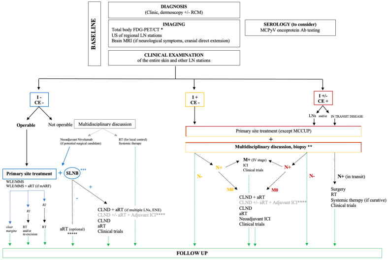
    1. Lugowska I., Becker J.C., Ascierto P.A., Veness M., Blom A., Lebbe C., Migliano E., Hamming-Vrieze O., Goebeler M., Kneitz H., et al. Merkel cell carcinoma: ESMO-EURACAN Clinical Practice Guideline for diagnosis, treatment and follow-up. ESMO Open. 2024;9:102977. doi: 10.1016/j.esmoop.2024.102977. - DOI - PMC - PubMed
    1. Gauci M.L., Aristei C., Becker J.C., Blom A., Bataille V., Dreno B., Del Marmol V., Forsea A.M., Fargnoli M.C., Grob J.J., et al. Diagnosis and treatment of Merkel cell carcinoma: European consensus-based interdisciplinary guideline—Update 2022. Eur. J. Cancer. 2022;171:203–231. doi: 10.1016/j.ejca.2022.03.043. - DOI - PubMed
    1. SEER*Explorer: An Interactive Website for SEER Cancer Statistics. [(accessed on 23 December 2024)]; Surveillance Research Program, National Cancer Institute. Available online: https://seer.cancer.gov/explorer/

LinkOut - more resources