. 2025 Jul 7;17(13):2266.
doi: 10.3390/cancers17132266.

Establishment of an Orthotopic and Metastatic Colorectal Cancer Mouse Model Using a Tissue Adhesive-Based Implantation Method

Affiliations

Establishment of an Orthotopic and Metastatic Colorectal Cancer Mouse Model Using a Tissue Adhesive-Based Implantation Method

Sang Bong Lee et al. Cancers (Basel). .

Abstract

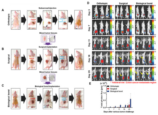
Background: To overcome the limitations of conventional CRC (colorectal cancer) mouse models in replicating metastasis and enabling efficient therapeutic evaluation, we developed a novel implantation method using tissue adhesive to establish reproducible orthotopic and metastatic tumors. Conventional models using injection or suturing techniques often suffer from technical complexity, inconsistent tumor establishment, and limited metastatic reliability. Methods: We developed and validated a novel orthotopic and metastatic CRC model utilizing tissue adhesive for tumor transplantation. Uniform tumor fragments derived from bioluminescent HCT116/Luc xenografts were affixed to the cecum of nude mice. Tumor growth and metastasis were monitored through bioluminescence imaging and confirmed by the results of histological analysis of metastatic lesions. The model's utility for therapeutic testing was evaluated using MK801, an NMDA receptor antagonist. Results: The biological-based model demonstrated rapid and reproducible tumor implantation (<5 min), consistent primary tumor growth, and robust metastasis to the liver and lungs. The biological-based approach achieved 80% tumor engraftment (4/5), with consistent metastasis to the liver and lungs in all mice, compared with lower and variable metastasis rates in injection (0%, 0/5) and suturing (20%, 1/5) methods. MK801 treatment significantly suppressed both primary tumor growth and metastasis, validating the model's suitability for preclinical drug evaluation. Conclusions: By enabling rapid, reproducible, and spontaneous formation of metastatic lesions using a minimally invasive tissue adhesive technique, our model represents a significant methodological advancement that supports high-throughput therapeutic screening and bridges the gap between experimental modeling and clinical relevance in colorectal cancer research.

Keywords: HCT116-Luc colon cancer cells; bioluminescence imaging; colorectal cancer; orthotopic model; preclinical drug evaluation; tissue adhesive (biological bond) implantation.


Conflict of interest statement

The authors declare that there are no conflicts of interest regarding the publication of this paper.

Figures

Figure 6
Figure 6
Evaluation of anticancer efficacy using biological bond- and surgery (suture)-based orthotopic colon cancer models. (A) In vivo bioluminescence imaging performed one day after tumor implantation confirmed successful model establishment in all groups. Mice were randomly assigned and treated with MK801 (1 mg/kg, intraperitoneally, daily on Days 1–5 post-implantation). Bioluminescent signals were monitored over 4 weeks. A reduction in tumor signal intensity was observed in both MK801-treated groups, while liver metastasis was only detected in the untreated biological bond group. (B) Quantification of total photon flux from the tumor regions at the final time point showed a consistent decrease in signal in the MK801-treated groups, supporting the therapeutic effect of NMDA receptor inhibition. The biological bond model also exhibited enhanced and reproducible tumor engraftment compared with the surgery-based model.
Figure 1
Figure 1
Schematic workflow for the development of a novel orthotopic and metastatic colorectal cancer model using biological tissue adhesive. The diagram illustrates: (1) subcutaneous xenograft formation using HCT116-Luc cells in nude mice; (2) harvesting and trimming of tumor tissue into standardized fragments; (3) three orthotopic implantation approaches direct injection into the cecum, surgical suturing of tumor fragments onto the cecum, and biological bonding; (4) closure of the abdominal incision using adhesive; (5) longitudinal monitoring of tumor growth and metastasis via bioluminescence imaging (IVIS); and (6) post-mortem detection of metastases in distant organs such as liver and lungs, indicated by corresponding anatomical icons.
Figure 2
Figure 2
Validation and preparation of subcutaneous xenograft tissue for orthotopic transplantation in luminescent colon cancer models. (A) Representative photographic and bioluminescent images of nude mice bearing subcutaneous HCT116-Luc tumors 7 days post-implantation. (B) Quantitative comparison of bioluminescent signal intensity between tumor and surrounding normal tissue, demonstrating tumor-specific signal. (C) Gross morphology of excised tumor tissues, trimmed to uniform sizes for transplantation. (D) Histogram showing tumor fragment weight consistency (mean ± SD, n = 5), confirming reproducibility of tissue preparation for orthotopic implantation.
Figure 3
Figure 3
Comparative evaluation of three orthotopic colon cancer implantation techniques. (A) Representative schematic and bioluminescent images of the orthotopic injection model, where tumor fragments or cell suspensions are delivered via syringe to the cecum wall. (B) Surgical suturing model, in which tumor fragments are physically attached to the cecal surface using suture threads. (C) Biological bonding model, where tumor fragments are affixed to the cecum using tissue adhesive for rapid and reproducible implantation. (D,E) Longitudinal quantification of total tumor photon flux (bioluminescence signal) from Day 3 to Day 25 post-implantation. The biological bonding group demonstrates higher tumor signal intensity and lower variability over time, compared with injection and suturing groups. Asterisks indicate statistically significant differences (p < 0.05).
Figure 4
Figure 4
(A) In vivo and ex vivo bioluminescence imaging of cecum (primary tumor site), liver, and lung tissues 25 days after tumor implantation via biological bonding. In vivo images show signal from luciferase-expressing HCT116/Luc colon cancer cells. Color bars represent radiance intensity (photons/sec/cm2/sr), with higher values indicating stronger bioluminescent signals associated with higher tumor burden. A consistent threshold of [state threshold if known, e.g., 1 × 10⁵ p/s/cm2/sr] was applied to define metastatic signals. (B) Representative gross anatomical and ex vivo IVIS images of resected livers showing metastatic lesions on the hepatic surface, with both ventral and dorsal views presented. Bioluminescent signal in the liver confirms metastatic spread from the cecum. These results collectively demonstrate the radiance-based detection of tumor progression from luciferase-tagged colorectal cancer xenografts.
Figure 5
Figure 5
Histological validation of metastatic colon cancer in major organs following biological bond-based tumor implantation. Representative H&E-stained tissue sections from (A) cecum, (B) liver, and (C) lung demonstrate the presence of metastatic tumor lesions (black arrows). Metastatic foci are characterized by dense clusters of atypical epithelial cells infiltrating normal tissue architecture. These findings confirm that bioluminescent signals detected by in vivo and ex vivo IVIS imaging correspond to actual metastatic colon cancer lesions in distant organs. The results support the biological fidelity of the model in replicating advanced-stage colorectal cancer metastasis.

Similar articles

References

    1. Chaffer C.L., Weinberg R.A. A perspective on cancer cell metastasis. Science. 2011;331:1559–1564. doi: 10.1126/science.1203543. - DOI - PubMed
    1. Valastyan S., Weinberg R.A. Tumor metastasis: Molecular insights and evolving paradigms. Cell. 2011;147:275–292. doi: 10.1016/j.cell.2011.09.024. - DOI - PMC - PubMed
    1. Gupta G.P., Massagué J. Cancer metastasis: Building a framework. Cell. 2006;127:679–695. doi: 10.1016/j.cell.2006.11.001. - DOI - PubMed
    1. Steeg P.S. Targeting metastasis. Nat. Rev. Cancer. 2016;16:201–218. doi: 10.1038/nrc.2016.25. - DOI - PMC - PubMed
    1. Ganesh K., Massagué J. Targeting metastatic cancer. Nat. Med. 2021;27:34–44. doi: 10.1038/s41591-020-01195-4. - DOI - PMC - PubMed

LinkOut - more resources