

Comparison of Dynamic Susceptibility Contrast and Arterial Spin Labeling MRI Perfusion in the Assessment of Stroke and Steno-Occlusive Disease: A Systematic Review and Meta-Analysis
- PMID: 40647577
- PMCID: PMC12249046
- DOI: 10.3390/diagnostics15131578
Comparison of Dynamic Susceptibility Contrast and Arterial Spin Labeling MRI Perfusion in the Assessment of Stroke and Steno-Occlusive Disease: A Systematic Review and Meta-Analysis
Abstract
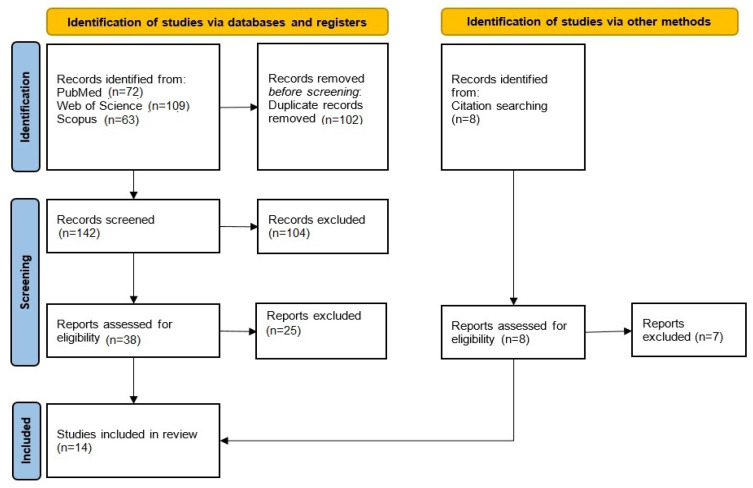
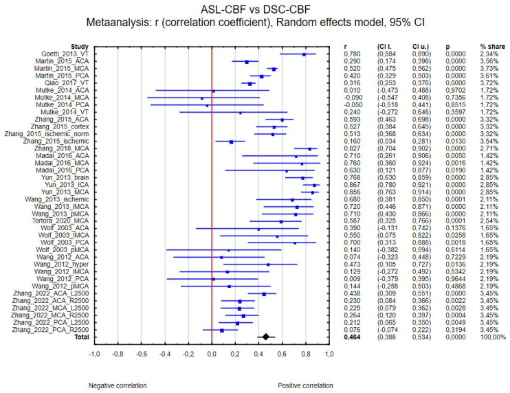
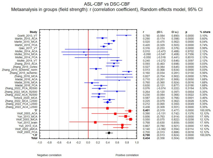
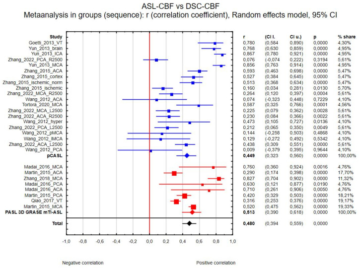
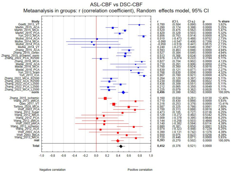
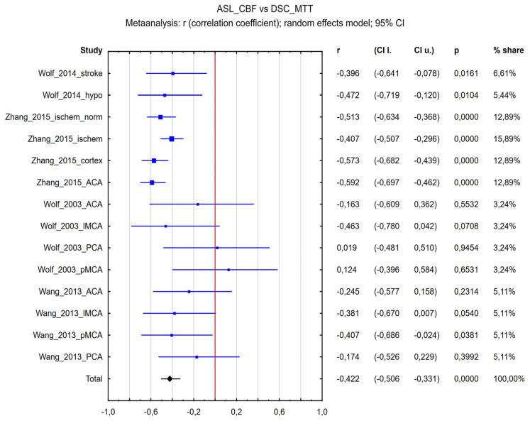
Background/Objectives: Assessment of the hemodynamic status of the brain in patients with cerebrovascular diseases is crucial for providing valuable clinical information. Various magnetic resonance perfusion sequences are used in studies, and one of the current challenges is comparing methods utilizing exogenous and endogenous contrast. This meta-analysis aimed to evaluate the correlation between arterial spin labeling (ASL)-derived perfusion parameters and those obtained by dynamic susceptibility contrast (DSC) perfusion in stroke and steno-occlusive diseases. Methods: A systematic review and meta-analysis were conducted, including 14 studies that reported correlation coefficients between perfusion MRI sequences in the assessment of stroke or steno-occlusive diseases. The correlation between ASL-derived cerebral blood flow (ASL-CBF) and DSC-derived cerebral blood flow (DSC-CBF) was analyzed, considering different magnetic field strengths (1.5 T and 3.0 T), sequence types, and brain regions. Additionally, real and normalized data were compared. Results: A moderate positive correlation was found between ASL-CBF and DSC-CBF (R = 0.464). Subgroup analysis demonstrated that ASL-CBF and DSC-CBF correlated at 3.0 T (R = 0.401) and 1.5 T (R = 0.700). No significant differences were observed in correlation coefficients based on sequence type or brain region. Normalized data demonstrated a higher correlation coefficient compared to real data (Rreal = 0.393, Rnorm = 0.496). Additionally, the correlation coefficient between ASL-CBF and DSC-derived mean transit time (DSC-MTT) for all included studies was R = -0.422. Conclusions: ASL-derived perfusion parameters demonstrate moderate-to-high agreement with DSC perfusion parameters in stroke and steno-occlusive patients. These findings support the potential utility of ASL as a non-invasive alternative to DSC perfusion imaging in clinical and research settings.
Keywords: arterial spin labeling; brain stroke; dynamic susceptibility contrast; perfusion imaging; steno-occlusive disease.
Conflict of interest statement
The authors declare no conflicts of interest.
Figures

References
-
- Weinreb J.C., Rodby R.A., Yee J., Wang C.L., Fine D., McDonald R.J., Perazella M.A., Dillman J.R., Davenport M.S. Use of Intravenous Gadolinium-Based Contrast Media in Patients with Kidney Disease: Consensus Statements from the American College of Radiology and the National Kidney Foundation. Radiology. 2021;298:28–35. doi: 10.1148/radiol.2020202903. - DOI - PubMed
-
- Levin J.M., Kaufman M.J., Ross M.H., Mendelson J.H., Maas L.C., Cohen B.M., Renshaw P.F. Sequential Dynamic Susceptibility Contrast MR Experiments in Human Brain: Residual Contrast Agent Effect, Steady State, and Hemodynamic Perturbation. Magn. Reson. Med. 1995;34:655–663. doi: 10.1002/mrm.1910340503. - DOI - PubMed
Publication types
LinkOut - more resources
Full Text Sources
