Learning Curve of Ultrasound-Guided Percutaneous Needle Biopsy for Pleural Lesions: A Retrospective Study at Two Tertiary Referral Hospitals
- PMID: 40647612
- PMCID: PMC12249106
- DOI: 10.3390/diagnostics15131613
Learning Curve of Ultrasound-Guided Percutaneous Needle Biopsy for Pleural Lesions: A Retrospective Study at Two Tertiary Referral Hospitals
Abstract
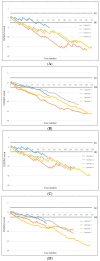
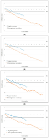
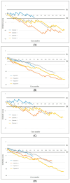
Background/Objectives: Ultrasound (US)-guided percutaneous pleural needle biopsy (PCPNB) is increasingly performed as a minimally invasive approach for the diagnosis of pleural lesions. However, no prior studies have investigated the learning curve for this method. The purpose of this study was to assess the learning curve of US-guided PCPNB using the cumulative summation (CUSUM) method and to calculate the number of procedures to achieve proficiency. Methods: A retrospective analysis was performed on 491 US-guided PCPNBs performed by four board-certified radiologists at two tertiary referral hospitals from January 2012 to December 2024. The standard and risk-adjusted CUSUM (RA-CUSUM) techniques were used to evaluate diagnostic success and false-negative rates. The potential impact of previous percutaneous needle biopsy (PCNB) experience on the learning curve was also assessed. Results: The overall diagnostic success and false-negative rate were 89.2% (438/491) and 8.1% (40/491), respectively. Operators achieved acceptable diagnostic success in US-guided PCPNB after a median of 15 (range, 12-45) procedures in standard CUSUM analysis and 22 (range, 10-33) procedures in RA-CUSUM analysis. Acceptable false-negative rates were attained after 18 (range, 14-42) and 24 (range, 12-44) procedures, respectively. Operators with prior PCNB experience required 12 procedures to achieve both acceptable diagnostic success and an acceptable false-negative rate. In contrast, those without experience required 29 and 27 procedures, respectively. Complications occurred in 1.4% (7/491), including one major complication (0.2%). Conclusions: Diagnostic proficiency and procedural safety in performing US-guided PCPNB improved with increasing operator experience. The low complication rate highlights the clinical safety and feasibility of US-guided PCPNB.
Keywords: learning curve; pleura; ultrasound-guided biopsy.
Conflict of interest statement
The authors have no financial conflicts of interest.
Figures


Similar articles
-
Comparison of Two Modern Survival Prediction Tools, SORG-MLA and METSSS, in Patients With Symptomatic Long-bone Metastases Who Underwent Local Treatment With Surgery Followed by Radiotherapy and With Radiotherapy Alone.Clin Orthop Relat Res. 2024 Dec 1;482(12):2193-2208. doi: 10.1097/CORR.0000000000003185. Epub 2024 Jul 23. Clin Orthop Relat Res. 2024. PMID: 39051924
-
Ultrasound guidance versus anatomical landmarks for internal jugular vein catheterization.Cochrane Database Syst Rev. 2015 Jan 9;1(1):CD006962. doi: 10.1002/14651858.CD006962.pub2. Cochrane Database Syst Rev. 2015. PMID: 25575244 Free PMC article.
-
Are Current Survival Prediction Tools Useful When Treating Subsequent Skeletal-related Events From Bone Metastases?Clin Orthop Relat Res. 2024 Sep 1;482(9):1710-1721. doi: 10.1097/CORR.0000000000003030. Epub 2024 Mar 22. Clin Orthop Relat Res. 2024. PMID: 38517402 Free PMC article.
-
Ultrasound guidance versus anatomical landmarks for subclavian or femoral vein catheterization.Cochrane Database Syst Rev. 2015 Jan 9;1(1):CD011447. doi: 10.1002/14651858.CD011447. Cochrane Database Syst Rev. 2015. PMID: 25575245 Free PMC article.
-
Ultrasound guidance for upper and lower limb blocks.Cochrane Database Syst Rev. 2015 Sep 11;2015(9):CD006459. doi: 10.1002/14651858.CD006459.pub3. Cochrane Database Syst Rev. 2015. PMID: 26361135 Free PMC article.
References
-
- Bødtger U., Halifax R.J. Pleural Disease. European Respiratory Society; Lausanne, Switzerland: 2020. Epidemiology: Why is pleural disease becoming more common? pp. 1–12.
-
- Boy D., Shaw J.A., Koegelenberg C.F.N. Ultrasound-guided pleural biopsy. Eurasian J. Pulmonol. 2023;25:1. doi: 10.14744/ejp.2021.9621. - DOI
LinkOut - more resources
Full Text Sources

