Review
. 2025 Jun 26;15(13):1625.
doi: 10.3390/diagnostics15131625.

MR Defecography Improves Diagnosis of Postoperative Pelvic Floor Dysfunction After Gynecological Surgery

Affiliations
Review

MR Defecography Improves Diagnosis of Postoperative Pelvic Floor Dysfunction After Gynecological Surgery

Rosa Alba Pugliesi et al. Diagnostics (Basel). .

Abstract

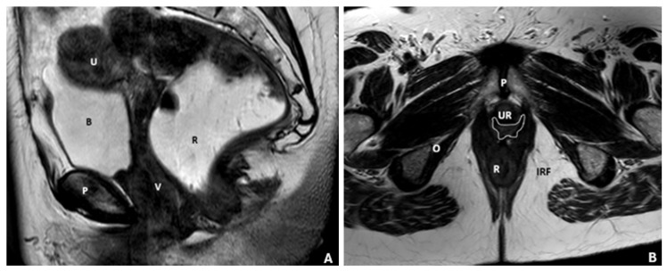
Pelvic floor dysfunction (PFD) is one of the most significant postoperative consequences in gynecological surgery, leading to impaired bowel function, structural alteration, and reduced quality of life. The conventional technique using fluoroscopic defecography and perineal ultrasonography provides an incomplete assessment of multi-compartment defects and post-surgical changes. Magnetic resonance defecography (MRD) represents a valuable alternative imaging method in the assessment of PFD following gynecological surgery, increasing diagnostic accuracy and enabling personalized treatment planning. MRD achieves high-resolution multi-compartmental assessment of the pelvic floor in dynamic states. Particularly, it is able to detect postoperative complications such as mesh retraction, organ prolapse, and fistula formation, not visible to other modalities. This narrative review discusses the role of MRD in diagnosing PFD and its advantages in detecting functional and anatomical changes following gynecological surgery. This review also examined the ability of MRD to demonstrate surgical changes and its contribution to possible standardization in clinical practice.

Keywords: MRI defecography; gynecological surgery; imaging techniques; pelvic floor dysfunction; pelvic organ prolapse; postoperative complications.


Conflict of interest statement

Roberto Cannella has the following disclosures, not related to this work: support for attending meetings from Bracco and Bayer; speaker for Bayer; had research collaboration with Siemens Healthineers. The other authors declare no conflicts of interest.

Figures

Figure 1
Figure 1
MRD of pelvic floor anatomy. (A) Sagittal T2-weighted turbo spin-echo (TSE) MRI illustrating normal female pelvic anatomy, clearly depicting the perineal body and levator plate. Anatomical landmarks include the bladder (B), uterus (U), vagina (V), rectum (R), and pubic bone (P). (B) Axial T2-weighted fast spin-echo (FSE) MRI demonstrating the pelvic floor anatomy. Identifiable structures include the pubic symphysis (P), rectus muscle (R), ischiorectal fossa (IRF), the normal butterfly-shaped vagina (outlined by a white line), urethra (UR), and the obturator muscle (O).
Figure 2
Figure 2
Female pelvic compartments. Sagittal T2-TSE image representing the three pelvic compartments: anterior, middle, and posterior. The anterior compartment, containing the bladder and urethra (U); the middle compartment, containing the uterus, cervix, and vagina (V); and the posterior compartment, containing the anus, anal canal, rectum (R), and sigmoid colon. A fourth “virtual” compartment called the cul-de-sac is also shown.
Figure 3
Figure 3
Pelvic muscles on axial MRI. Axial T2-weighted images (AC) demonstrate normal pelvic diaphragm anatomy, including the iliococcygeus, ischiococcygeus, puborectalis (*), and obturator internus muscles. C = coccyx, P = pubic symphysis, R = rectum, U = urethra, B = bladder, V = vagina.
Figure 4
Figure 4
Normal female pelvic floor anatomy on axial MRI. Axial T2-weighted image shows the urogenital hiatus (white line) and key urethral support structures, including periurethral (arrow), paraurethral (arrowhead), and pubourethral (star) ligaments, with characteristic H-shaped vagina (V), urethra (U), pubic symphysis (P), and rectum (R).
Figure 5
Figure 5
MR defecography in a post-hysterectomy patient. (A) Sagittal T2-weighted turbo spin-echo (TSE) MRI illustrating the female pelvic anatomy at rest; (B) sagittal balanced turbo field echo (BTFE) sequence during the evacuation phase; (C) axial T2-weighted turbo spin-echo (TSE) sequence showing the pelvic floor anatomy. Identifiable structures include the pubic symphysis (P), rectum (R), ischiorectal fossa (IRF), bladder (B), and the obturator muscle (O).
Figure 6
Figure 6
Normal position at rest (A), during straining (B), and defecation (C). There is a mild descent of all the three compartments (urinary bladder, vaginal vault, and anorectal junction). Identifiable structures include the pubic symphysis (P), rectum (R), bladder (B), and uterus (U).
Figure 7
Figure 7
Sagittal T2-weighted MR defecography images demonstrate anorectal angle dynamics. (A) At rest post-rectal gel filling, the angle measures 81°, with the anorectal junction at the pubococcygeal line (PCL). (B) During pre-defecation contraction, the angle narrows to 54°. (C) During defecation straining, rectal prolapse widens the angle to 120°, with a 6 cm anorectal junction descent. Identifiable structures include the pubic symphysis (P), rectum (R), bladder (B), and uterus (U).
Figure 8
Figure 8
Sagittal T2-weighted MR defecography at squeeze shows normal pelvic anatomy. The pubococcyxgeal line (PCL), H line, M line, and mid-pubic line (MPL) are displayed. The anorectal junction aligns with the PCL, the H and M lines measure within normal limits, and no pelvic floor descent or organ prolapse is observed. Identifiable structures include the pubic symphysis (P), rectum (R), bladder (B), and uterus (U).
Figure 9
Figure 9
Sagittal midline images pre-evacuation (A) and during evacuation (B) illustrate descending perineal syndrome with tricompartimental prolapse—cystocele (star), uterine, and rectal prolapse—alongside anterior rectocele and rectal mucosa intussusception (white arrow). The evacuation phase is essential for revealing the full extent of pelvic floor dysfunction. M-line (dark) and PCL (white) are shown. Identifiable structures include the pubic symphysis (P), rectum (R), bladder (B), and uterus (U).
Figure 10
Figure 10
At rest (A), pelvic organs maintain normal positions. During defecation (B), notable descent of the peritoneal sac with small bowel loops defines an enterocele (white dotted). Associated findings include bladder and anorectal descent, anterior rectocele, rectorectal intussusception, cystocele, hysterocele, and severe elitrocele. M-line (orange) and PCL (white) are indicated. Identifiable structures include the pubic symphysis (P), rectum (R), bladder (B), and uterus (U).
Figure 11
Figure 11
Sagittal midline image of the anterior/posterior rectocele during evacuation, illustrating both anterior and posterior rectoceles. The anorectal wall appears normal at rest (A), but during evacuation (B) the white dotted lines highlight the extent of the rectocele—40 mm anteriorly and 13 mm posteriorly. The PCL (white continuous), and anterior/posterior rectocele (white dotted) are depicted. Identifiable structures include the pubic symphysis (P), rectum (R), bladder (B), and uterus (U).
Figure 12
Figure 12
MRI at rest (A) and during defecation (B) showing isthmocele, cystocele, and anterior rectocele. The M line (dark continuous) and PCL (white continuous) are marked. The cystocele and anterior isthmocele are indicated by white dotted lines, while the anterior rectocele is highlighted with black dotted lines. Identifiable structures include the pubic symphysis (P), rectum (R), bladder (B), and uterus (U).

Similar articles

References

    1. Peinado-Molina R.A., Hernández-Martínez A., Martínez-Vázquez S., Rodríguez-Almagro J., Martínez-Galiano J.M. Pelvic floor dysfunction: Prevalence and associated factors. BMC Public Health. 2023;23:2005. doi: 10.1186/s12889-023-16901-3. - DOI - PMC - PubMed
    1. Tim S., Mazur-Bialy A.I. The most common functional disorders and factors affecting female pelvic floor. Life. 2021;11:1397. doi: 10.3390/life11121397. - DOI - PMC - PubMed
    1. Al-Badr A., Saleem Z., Kaddour O., Almosaieed B., Dawood A., Al-Tannir M., AlTurki F., Alharbi R., Alsanea N. Prevalence of pelvic floor dysfunction: A Saudi national survey. BMC Womens Health. 2022;22:27. doi: 10.1186/s12905-022-01609-0. - DOI - PMC - PubMed
    1. Benti Terefe A., Gemeda Gudeta T., Teferi Mengistu G., Abebe S.S. Determinants of pelvic floor disorders among women visiting the gynecology outpatient department in wolkite university specialized center, wolkite. Ethiop. Obs. Gynecol. Int. 2022;13:6949700. doi: 10.1155/2022/6949700. - DOI - PMC - PubMed
    1. Ferrari L., Gala T., Igualada-Martinez P., Brown H.W., Weinstein M., Hainsworth A. Multidisciplinary team (MDT) approach to pelvic floor disorders. Continence. 2023;7:100716. doi: 10.1016/j.cont.2023.100716. - DOI

LinkOut - more resources