Review
. 2025 Jul 1;15(13):1680.
doi: 10.3390/diagnostics15131680.

Expanding Role of Contrast-Enhanced Ultrasound and Elastography in the Evaluation of Abdominal Pathologies in Children

Affiliations
Review

Expanding Role of Contrast-Enhanced Ultrasound and Elastography in the Evaluation of Abdominal Pathologies in Children

Natae Fekadu Lemessa et al. Diagnostics (Basel). .

Abstract

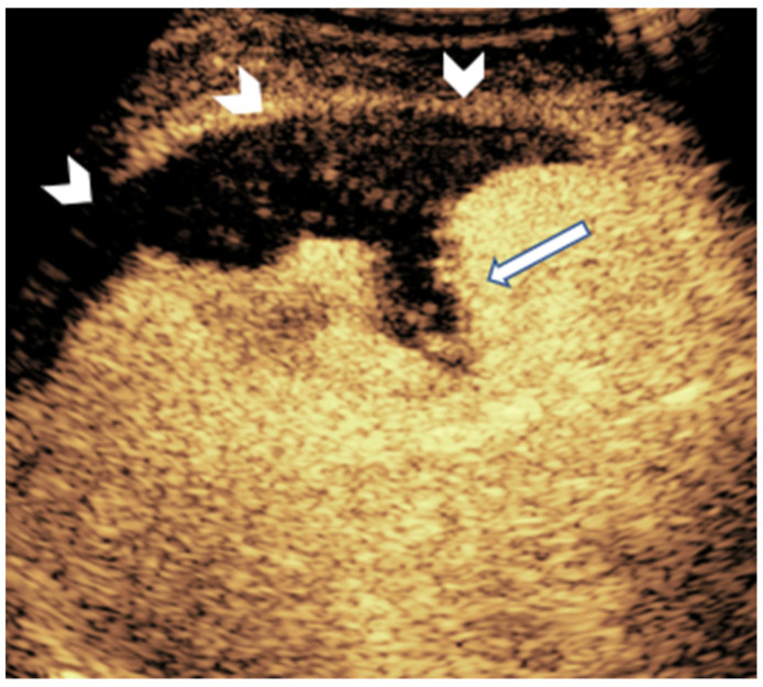
Contrast-enhanced ultrasound and elastography are two ultrasound technologies that are becoming increasingly popular in the evaluation of different abdominal pathologies in children. The use of these technologies has expanded the diagnostic scope of ultrasound into areas that were traditionally covered by advanced imaging modalities such as computed tomography, magnetic resonance imaging, and fluoroscopy. In this review, we summarize the use of contrast-enhanced ultrasound and elastography in the evaluation of hepatic, renal, pancreatic, splenic, urinary tract, and scrotal pathologies in children. We describe the technical aspects, applications, and limitations, intending to make readers more acquainted with the technologies.

Keywords: abdominal imaging; contrast-enhanced ultrasound; elastography; multiparametric ultrasound; pediatrics.


Conflict of interest statement

The authors declare no conflict of interest.

Figures

Figure 1
Figure 1
CEUS in a 24-day-old girl with hemangioma. (a) A transverse scan of the right lobe of the liver shows multiple hypoechoic masses (arrow). (b) Arterial phase shows nodular peripheral hyperenhancement. (c) Porto-venous phase shows hyperenhancement with progressive centripetal filling. (d) Delayed phase shows the absence of rapid washout.
Figure 2
Figure 2
CEUS in a 16-year-old girl with hepatoblastoma. (a) A longitudinal scan of the liver shows a well-defined, homogenously hypoechoic mass in grayscale (arrow). (b) Iso- to slightly hyperintense enhancement on early arterial phase. (c) Hypoenhancement in porto-venous phase. (d) Early washout on delayed phase indicative of the malignant nature of the mass.
Figure 3
Figure 3
Ileocolonic Crohn’s disease in a 13-year-old boy. Dual-screen mode with simultaneous display of grayscale (left) and contrast-enhanced (right) proximal ascending colon in the longitudinal plane shows smoothly thickened proximal ascending colon with rapid wash-in and avid enhancement (arrows). The enhancement persisted in porto-venous and delayed phases.
Figure 4
Figure 4
An 11-year-old girl who had recently undergone left lower renal subsegmental artery embolization. Post-procedure contrast-enhanced renal ultrasound shows a lack of parenchymal enhancement in the lowermost portion of the left kidney (arrow), suggestive of the absence of perfusion.
Figure 5
Figure 5
Gray scale (left) and contrast-enhanced (right) pancreatic ultrasound shows enlarged and heterogeneous pancreas (arrowhead) and non-enhancement of the almost entire pancreatic parenchyma (arrow), revealing extensive necrosis in a case of acute pancreatitis [61].
Figure 6
Figure 6
Contrast-enhanced urosonographic staging of vesicoureteric reflux. Images acquired from different children with varying degrees of reflux. (a) Grade I reflux: Contrast limited to the distal ureter. (b) Grade II reflux: Contrast extending to a non-dilated pelvicalyceal system. (c) Grade III reflux: Contrast seen in mildly dilated pelvicalyceal system with preserved calyceal contour. (d) Grade IV reflux: Contrast in moderately dilated pelvicalyceal system with blunted fornices but preserved papillary impression. (e) Grade V reflux: Contrast seen in severely dilated collecting system with fornices blunted and lost papillary impression. Black star: contrast filled bladder. White arrow: distal ureter.
Figure 7
Figure 7
A contrast-enhanced splenic ultrasound in a 9-year-old boy with a history of abdominal trauma shows non-enhancing parenchymal (arrow) and subcapsular (arrowhead) areas representing parenchymal laceration and subcapsular hematoma, respectively.
Figure 8
Figure 8
A 16-year-old-girl with acute myelogenous leukemia and multiorgan failure developed a peripheral, wedge-shaped, non-enhancing splenic lesion (arrow) on CEUS, likely denoting splenic infarct.
Figure 9
Figure 9
CEUS of the spleen in a 15-year-old boy with hepatosplenic candidiasis shows multiple non-enhancing nodules (arrow) suggestive of microabscesses.
Figure 10
Figure 10
Normal grayscale and enhancement pattern of the testis in an 11-year-old boy. (a) Grayscale. (b) Arterial phase showing early linear arterial enhancement. (c) Parenchymal phase showing homogenous testicular enhancement. (d) Gradual washout.
Figure 11
Figure 11
Congenital hepatic fibrosis in a 5-year-old girl. Mean shear wave elastography at the right lobe of the liver measured 3.9 m/s. The normal pediatric liver stiffness value using this ultrasound scanner and technique is approximately 1.16 m/s (SD +/− 0.14 m/s). Therefore, the finding is suggestive of fibrosis.

Similar articles

References

    1. Dillman J.R., Gee M.S., Ward C.G., Drum E.T., States L.J. Imaging sedation and anesthesia practice patterns in pediatric radiology departments—A survey of the Society of Chiefs of Radiology at Children’s Hospitals (SCORCH) Pediatr. Radiol. 2021;51:1497–1502. doi: 10.1007/s00247-021-04996-y. - DOI - PubMed
    1. Ntoulia A., Anupindi S.A., Back S.J., Didier R.A., Hwang M., Johnson A.M., Sommer F.G., Bellah R.D., Biko D.M., Darge K., et al. Contrast-enhanced ultrasound: A comprehensive review of safety in children. Pediatr. Radiol. 2021;51:2161–2180. doi: 10.1007/s00247-021-05223-4. - DOI - PMC - PubMed
    1. Ajmal S. Contrast-Enhanced Ultrasonography: Review and Applications. Cureus. 2021;13:e18243. doi: 10.7759/cureus.18243. - DOI - PMC - PubMed
    1. Takahashi M.S., Yamanari M.G.I., Suzuki L., Pedrosa É.F.N.C., Lopes R.I., Chammas M.C. Use of contrast-enhanced ultrasound in pediatrics. Radiol. Bras. 2021;54:321–328. doi: 10.1590/0100-3984.2020.0167. - DOI - PMC - PubMed
    1. Squires J.H., McCarville M.B. Contrast-Enhanced Ultrasound in Children: Implementation and Key Diagnostic Applications. AJR Am. J. Roentgenol. 2021;217:1217–1231. doi: 10.2214/AJR.21.25713. - DOI - PubMed

LinkOut - more resources