

Deep Learning-Enhanced T1-Weighted Imaging for Breast MRI at 1.5T
- PMID: 40647680
- PMCID: PMC12248570
- DOI: 10.3390/diagnostics15131681
Deep Learning-Enhanced T1-Weighted Imaging for Breast MRI at 1.5T
Abstract
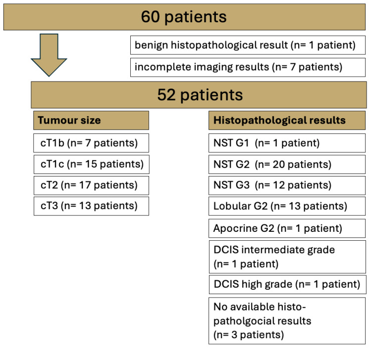
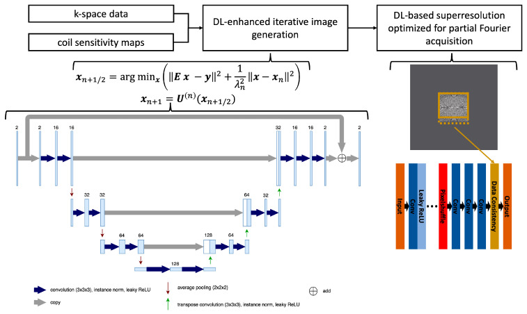
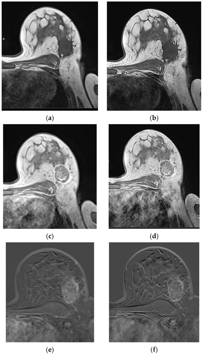
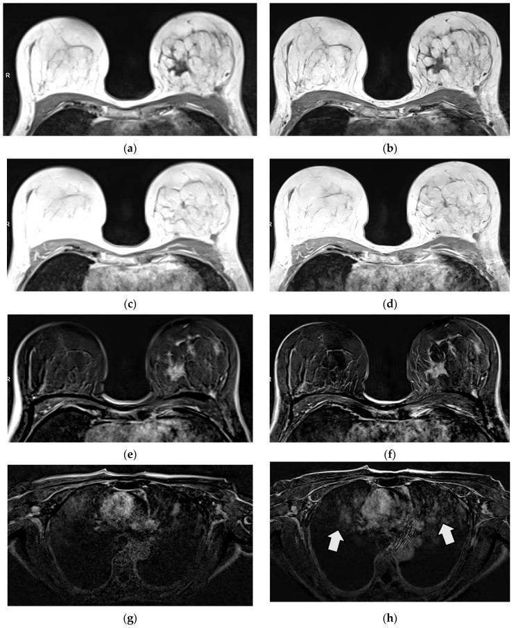
Background/Objectives: Assessment of a novel deep-learning (DL)-based T1w volumetric interpolated breath-hold (VIBEDL) sequence in breast MRI in comparison with standard VIBE (VIBEStd) for image quality evaluation. Methods: Prospective study of 52 breast cancer patients examined at 1.5T breast MRI with T1w VIBEStd and T1 VIBEDL sequence. T1w VIBEDL was integrated as an additional early non-contrast and a delayed post-contrast scan. Two radiologists independently scored T1w VIBE Std/DL sequences both pre- and post-contrast and their calculated subtractions (SUBs) for image quality, sharpness, (motion)-artifacts, perceived signal-to-noise and diagnostic confidence with a Likert-scale from 1: Non-diagnostic to 5: Excellent. Lesion diameter was evaluated on the SUB for T1w VIBEStd/DL. All lesions were visually evaluated in T1w VIBEStd/DL pre- and post-contrast and their subtractions. Statistics included correlation analyses and paired t-tests. Results: Significantly higher Likert scale values were detected in the pre-contrast T1w VIBEDL compared to the T1w VIBEStd for image quality (each p < 0.001), image sharpness (p < 0.001), SNR (p < 0.001), and diagnostic confidence (p < 0.010). Significantly higher values for image quality (p < 0.001 in each case), image sharpness (p < 0.001), SNR (p < 0.001), and artifacts (p < 0.001) were detected in the post-contrast T1w VIBEDL and in the SUB. SUBDL provided superior diagnostic certainty compared to SUBStd in one reader (p = 0.083 or p = 0.004). Conclusions: Deep learning-enhanced T1w VIBEDL at 1.5T breast MRI offers superior image quality compared to T1w VIBEStd.
Keywords: MRI; breast cancer; deep learning; diagnostic imaging.
Conflict of interest statement
Authors Marcel Dominik Nickel and Elisabeth Weiland were employed by the company Siemens Healthineers AG, Forchheim, Germany. The remaining authors declare that the research was conducted in the absence of any commercial or financial relationships that could be construed as a potential conflict of interest.
Figures
Similar articles
-
Optimizing contrast-enhanced abdominal MRI: A comparative study of deep learning and standard VIBE techniques.Clin Imaging. 2025 Aug 7;126:110581. doi: 10.1016/j.clinimag.2025.110581. Online ahead of print. Clin Imaging. 2025. PMID: 40795789
-
AI-Assisted Post Contrast Brain MRI: Eighty Percent Reduction in Contrast Dose.Acad Radiol. 2025 Aug;32(8):4767-4776. doi: 10.1016/j.acra.2024.10.026. Epub 2024 Nov 26. Acad Radiol. 2025. PMID: 39592383
-
Deep learning reconstruction for improving image quality of pediatric abdomen MRI using a 3D T1 fast spoiled gradient echo acquisition.Pediatr Radiol. 2025 Jul 18. doi: 10.1007/s00247-025-06313-3. Online ahead of print. Pediatr Radiol. 2025. PMID: 40679617
-
A systematic review of evidence on malignant spinal metastases: natural history and technologies for identifying patients at high risk of vertebral fracture and spinal cord compression.Health Technol Assess. 2013 Sep;17(42):1-274. doi: 10.3310/hta17420. Health Technol Assess. 2013. PMID: 24070110 Free PMC article.
-
Systemic pharmacological treatments for chronic plaque psoriasis: a network meta-analysis.Cochrane Database Syst Rev. 2017 Dec 22;12(12):CD011535. doi: 10.1002/14651858.CD011535.pub2. Cochrane Database Syst Rev. 2017. Update in: Cochrane Database Syst Rev. 2020 Jan 9;1:CD011535. doi: 10.1002/14651858.CD011535.pub3. PMID: 29271481 Free PMC article. Updated.
References
LinkOut - more resources
Full Text Sources
Research Materials
