

Association Between Microcalcification Patterns in Mammography and Breast Tumors in Comparison to Histopathological Examinations
- PMID: 40647686
- PMCID: PMC12248651
- DOI: 10.3390/diagnostics15131687
Association Between Microcalcification Patterns in Mammography and Breast Tumors in Comparison to Histopathological Examinations
Abstract
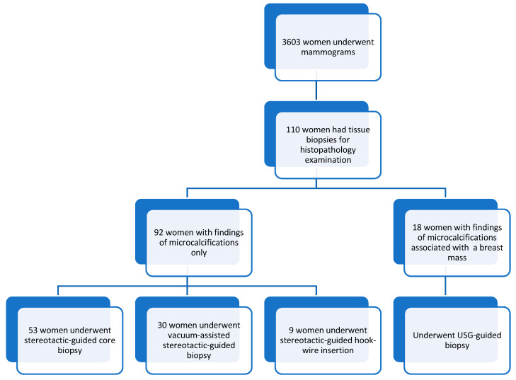
Background/Objectives: Accurately correlating mammographic findings with corresponding histopathologic features is considered one of the essential aspects of mammographic evaluation, guiding the next steps in cancer management and preventing overdiagnosis. The objective of this study was to evaluate patterns of mammographic microcalcifications and their association with histopathological findings related to various breast tumors. Methods: 110 out of 3603 women had microcalcification of BIRADS 3 or higher and were subjected to stereotactic/ultrasound (USG) guided biopsies, and hook-wire localization excision procedures. Ultrasound and mammography images were reviewed by experienced radiologists using the standard American College of Radiology Breast-Imaging Reporting and Data System (ACR BI-RADS). Results: Our study showed that features with a high positive predictive value (PPV) of breast malignancy were heterogeneous (75%), fine linear/branching pleomorphic microcalcifications (66.7%), linear (100%), and segmental distributions (57.1%). Features that showed a higher risk of association with ductal carcinoma in situ (DCIS) were fine linear/branching pleomorphic (odds ratio (OR): 3.952), heterogeneous microcalcifications (OR: 3.818), segmental (OR: 5.533), linear (OR: 3.696), and regional (OR: 2.929) distributions. Furthermore, the features with higher risks associated with invasive carcinoma had heterogeneous (OR: 2.022), fine linear/branching pleomorphic (OR: 1.187) microcalcifications, linear (OR: 6.2), and regional (OR: 2.543) distributions. The features of associated masses in mammograms that showed a high PPV of malignancy had high density (75%), microlobulation (100%), and spiculated margins (75%). Conclusions: We concluded that specific patterns and distributions of microcalcifications were indeed associated with a higher risk of malignancy. Those with fine linear or branching pleomorphic and segmental distribution were at a higher risk of DCIS, whereas those with heterogeneous morphology with a linear distribution were at a higher risk of invasive carcinoma.
Keywords: breast malignancy; ductal carcinoma in situ; histopathology; invasive carcinoma; mammogram; mass; microcalcifications.
Conflict of interest statement
The authors declare no conflicts of interest.
Figures
Similar articles
-
Mammographic density, endocrine therapy and breast cancer risk: a prognostic and predictive biomarker review.Cochrane Database Syst Rev. 2021 Oct 26;10(10):CD013091. doi: 10.1002/14651858.CD013091.pub2. Cochrane Database Syst Rev. 2021. PMID: 34697802 Free PMC article.
-
Early detection of breast cancer: benefits and risks of supplemental breast ultrasound in asymptomatic women with mammographically dense breast tissue. A systematic review.BMC Cancer. 2009 Sep 20;9:335. doi: 10.1186/1471-2407-9-335. BMC Cancer. 2009. PMID: 19765317 Free PMC article.
-
An Analysis of the Results of Breast Imaging Reporting and Data System 4 Lesions in Tertiary Care Center in South India.Indian J Surg Oncol. 2025 Feb;16(1):312-319. doi: 10.1007/s13193-024-02089-4. Epub 2024 Sep 12. Indian J Surg Oncol. 2025. PMID: 40114879
-
Can Mammography and Magnetic Resonance Imaging Predict the Preoperative Size and Nuclear Grade of Pure Ductal Carcinoma In Situ?Diagnostics (Basel). 2025 Jul 17;15(14):1801. doi: 10.3390/diagnostics15141801. Diagnostics (Basel). 2025. PMID: 40722550 Free PMC article.
-
Localization techniques for guided surgical excision of non-palpable breast lesions.Cochrane Database Syst Rev. 2015 Dec 31;2015(12):CD009206. doi: 10.1002/14651858.CD009206.pub2. Cochrane Database Syst Rev. 2015. PMID: 26718728 Free PMC article.
References
-
- Azizah A.M., Hashimah B., Nirmal K., Siti Zubaidah A.R., Puteri N.A., Nabihah A., Sukumaran R., Balqis B., Nadia S.M.R., Sharifah S.S.S., et al. Malaysia National Cancer Registry Report, 2012–2016. National Cancer Institute, Ministry of Health; Putrajaya, Malaysia: 2019.
-
- Gavric Z. Quality of Life of Women with Breast Cancer-Emotional and Social Aspects. Am. J. Cancer Prev. 2015;3:13–18.
LinkOut - more resources
Full Text Sources
