. 2025 Jun 26;17(13):1768.
doi: 10.3390/polym17131768.

Graphene Oxide-Enriched Polymer: Impact on Dental Pulp Cell Viability and Differentiation

Affiliations

Graphene Oxide-Enriched Polymer: Impact on Dental Pulp Cell Viability and Differentiation

Magdalena Vega-Quiroz et al. Polymers (Basel). .

Abstract

Background: Reconstructing maxillofacial defects is important in dentistry, so efforts are being made to develop materials that promote cell migration and repair. Graphene oxide (GO) is used to enhance the biocompatibility of polymethylmethacrylate (PMMA) due to its nanostructure.

Objective: to assess cytotoxicity, cell proliferation, and differentiation of human dental pulp stem cells (hDPSC) in response to a conventional PMMA (PMMA) and polymer enriched with GO (PMMA+GO).

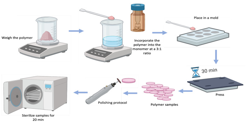
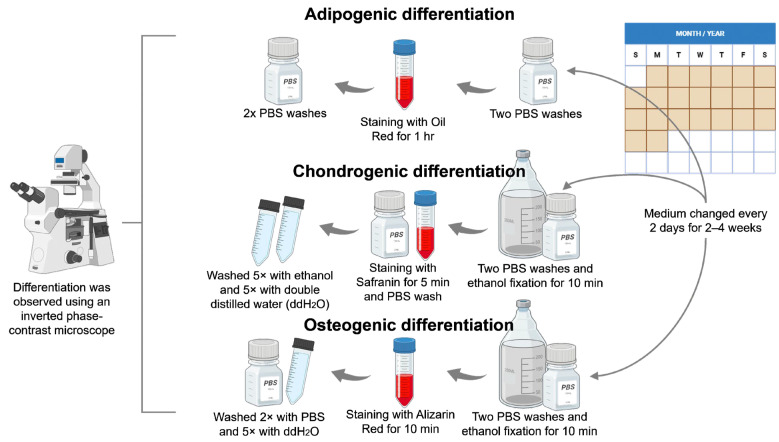
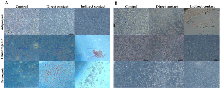
Methods: Experiments were carried out with primary hDPSC subcultures. The PMMA and PMMA+GO were tested in direct and indirect contact. Cytotoxicity (1 day) and proliferation (3, 7, and 14 days) were evaluated with an MTT bioassay. The osteogenic, adipogenic, and chondrogenic aspects were determinate with alizarin red, oil red, and safranine. Mean values, standard deviation, and percentages were calculated; data were analyzed with Shapiro-Wilks normality and Student's t-test.

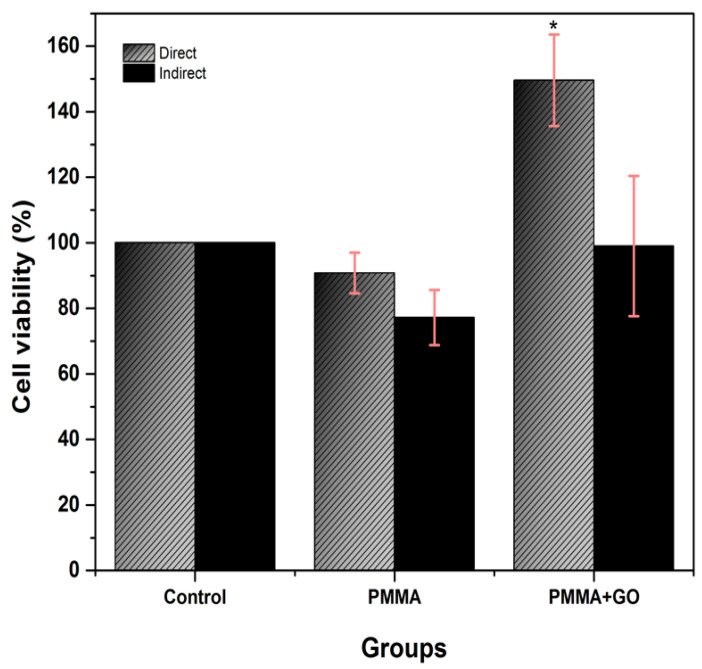
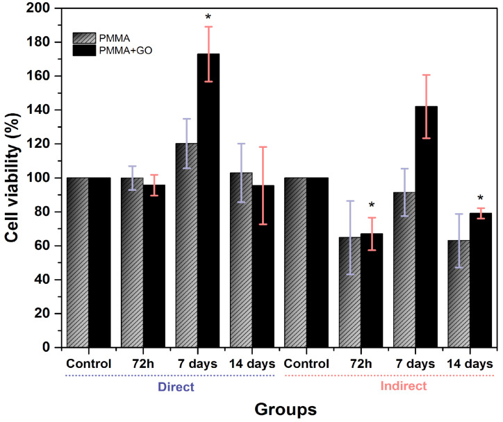
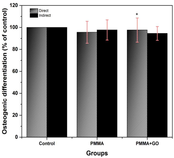
Results: The cell viability of PMMA and PMMA+GO in direct contact correspond to 90.8 ± 6.2, 149.6 ± 14.5 (1 day); 99.9 ± 7.0, 95.7 ± 6.1 (3 days); 120.2 ± 14.6, 172.9 ± 16.2 (7 days); and 102.9 ± 17.3, 95.4 ± 22.8 (14 days). For indirect contact, 77.2 ± 8.4, 99 ± 21.4 (1 day); 64.8 ± 21.6, 67.0 ± 9.6 (3 days); 91.4 ± 16.5, 142 ± 18.7 (7 days); and 63 ± 15.8, 79.1 ± 3.1 (14 days). PMMA+GO samples showed enhanced adipogenic, chondrogenic, and osteogenic aspects.

Conclusions: The integration of GO into PMMA biopolymers stimulates cell proliferation and differentiation, holding great promise for future applications in the field of biomedicine.

Keywords: cell proliferation; cytotoxicity; graphene oxide; polymethylmethacrylate.


Conflict of interest statement

Authors Dr. Hector Guzman-Juarez and Dr. Carlos Andres Alvarez-Gayoso are employed by the company Osforma. The remaining authors declare that the research was conducted in the absence of any commercial or financial relationships that could be construed as a potential conflict of interest.

Figures

Figure 1
Figure 1
Processing of conventional and GO polymer samples. Source: Direct obtained with BioRender 2025.
Figure 2
Figure 2
Schematic representation of the method for cell differentiation of hDPSC into adipogenic, chondrogenic, and osteogenic lineage. Source: Direct obtained by BioRender 2025.
Figure 3
Figure 3
PMMA+GO samples analyzed under an optical microscope, showing spherical particles (A); in the SEM photomicrograph, the lamellar morphology of GO is observed (B).
Figure 4
Figure 4
hDPSC cell cytotoxicity in direct and indirect 24 h contact with PMMA and PMMA+GO. Optical density 570 nm. Abs. range (0.137–3.303). Each bar represents the mean percentage ± standard deviation (SD). Statistical analysis was performed using one-way ANOVA, followed by Tukey’s post hoc test. Asterisks (*) indicate concentrations with statistically significant differences compared to the control group (p < 0.001, n = 9). Source: Direct.
Figure 5
Figure 5
hDPSC cell viability in 72 h, 7 days and 14 days direct and indirect contact with PMMA and PMMA. Optical density 570 nm. Abs. range (0.099–3.214). Each bar represents the mean percentage ± standard deviation (SD). Statistical analysis was performed using one-way ANOVA, followed by Tukey’s post hoc test. Asterisks (*) indicate concentrations with statistically significant differences compared to the control group (p < 0.001, n = 9). Source: Direct.
Figure 6
Figure 6
Comparative photomicrographs of hDPSC differentiation into adipogenic, chondrogenic, and osteogenic lineages under control conditions, direct contact, and indirect contact with conventional PMMA (A) and GO-enriched PMMA (B). Morphological evidence of lineage-specific differentiation is visible in each condition. Source: Direct observation.
Figure 7
Figure 7
Osteogenic differentiation in direct and indirect contact with PMMA and PMMA. Optical density 570 nm. Abs. range (0.045–0.052), ach value in the graph represents percentage of the mean and SD. One-way ANOVA was performed, Tukey’s post hoc, (*) represents concentrations with significant difference. Data represent mean and variance. ANOVA test * p < 0.05, Tukey’s post hoc, n = 9. Source: Direct.

Similar articles

References

    1. Brown P.T., Handorf A.M., Jeon W.B., Li W.J. Stem cell-based tissue engineering approaches for musculoskeletal regeneration. Curr. Pharm. Des. 2013;19:3429–3445. doi: 10.2174/13816128113199990350. - DOI - PMC - PubMed
    1. Umapathy V.R., Natarajan P.M., Swamikannu B. Regenerative Strategies in Dentistry: Harnessing Stem Cells, Biomaterials and Bioactive Materials for Tissue Repair. Biomolecules. 2025;15:546. doi: 10.3390/biom15040546. - DOI - PMC - PubMed
    1. Hynds R.E., Magin C.M., Ikonomou L., Aschner Y., Beers M.F., Burgess J.K., Heise R.L., Hume P.S., Krasnodembskaya A.D., Mei S.H., et al. Stem cells, cell therapies, and bioengineering in lung biology and diseases 2023. Am. J. Physiol. Lung Cell Mol. Physiol. 2024;327:L327–L340. doi: 10.1152/ajplung.00052.2024. - DOI - PMC - PubMed
    1. Bakhshandeh B., Ranjbar N., Abbasi A., Amiri E., Abedi A., Mehrabi M.R., Dehghani Z., Pennisi C.P. Recent progress in the manipulation of biochemical and biophysical cues for engineering functional tissues. Bioeng. Transl. Med. 2022;8:e10383. doi: 10.1002/btm2.10383. - DOI - PMC - PubMed
    1. Al-Rawe R.A., Al-Rammahi H.M., Cahyanto A., Ma’amor A., Liew Y.M., Sukumaran P., Wan Hassan W.N. Cuttlefish-Bone-Derived Biomaterials in Regenerative Medicine, Dentistry, and Tissue Engineering: A Systematic Review. J. Funct. Biomater. 2024;15:219. doi: 10.3390/jfb15080219. - DOI - PMC - PubMed

LinkOut - more resources