. 2025 Jun 26;14(13):1952.
doi: 10.3390/plants14131952.

Phylogenetic and Structural Insights into Melatonin Receptors in Plants: Case Study in Capsicum chinense Jacq

Affiliations

Phylogenetic and Structural Insights into Melatonin Receptors in Plants: Case Study in Capsicum chinense Jacq

Adrian Toledo-Castiñeira et al. Plants (Basel). .

Abstract

Recently, it has been proposed that plant melatonin receptors belong to the superfamily of G protein-coupled receptors (GPCRs). However, a detailed description of the phylogeny, protein structure, and binding properties of melatonin, which is still lacking, can help determine the signaling and function of this compound. Melatonin receptor homologs (PMTRs) were identified in 90 Viridiplantae sensu lato proteomes using profile Hidden Markov Models (HMM), which yielded 174 receptors across 87 species. Phylogenetic analysis revealed an expansion of PMTR sequences in angiosperms, which were grouped into three clades. Docking studies uncovered a conserved internal melatonin-binding site in PMTRs, which was analogous to the site in human MT1 receptors. Binding affinity simulations indicated this internal site exhibits stronger melatonin binding compared to a previously reported superficial pocket. Ligand-receptor interaction analysis and alanine scanning highlighted a major role of hydrophobic interactions, with hydrogen bonds contributing predominantly at the internal site, while non-interacting charged residues stabilize the binding pocket. Tunnel and ligand transport simulations suggested melatonin moves favorably through the internal cavity to access the binding site. Also, we presented for the first time details of these pockets in a non-model species, Capsicum chinense. Taken together, the structural analyses presented here illustrate opportunities and theoretical evidence for performing structure-function studies via mutations in specific residues within the proposed new melatonin-binding site in PMTRs, shedding light on their role in plant melatonin signaling.

Keywords: Arabidopsis; Capsicum; G protein-coupled receptor; HMM; melatonin; molecular docking; phylogeny; phytomelatonin receptor PMTR.


Conflict of interest statement

The authors declare no conflicts of interest.

Figures
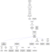
Figure 1
Figure 1
Graphical summary of the workflow followed in this study. Species spanning diverse taxonomic groups were selected based on León-García et al. [26] to build the HMM.
Figure 2
Figure 2
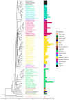
Evolutionary tree of the species included in this study (left). A consensus-rooted species tree was inferred by Orthofinder [27] from 90 plant proteomes. Horizontal bars (right) represent the number of sequences per species from a total of 174 sequences identified after the screening process.
Figure 3
Figure 3
Phylogenetic tree of putative melatonin receptors for 87 species across 39 plant families. Numbers along branches represent support values obtained by 1000 bootstraps. Angiosperm representatives were grouped into three clades, which are highlighted. Taxa groups were mapped into colors. Images representing well-known taxa group representatives were included. Protein topologies obtained with Tmbed [28] are represented on the right side of the tree, with transmembrane regions represented as dark blue bars. Red dots in the branch tips represent those melatonin receptors previously reported.
Figure 4
Figure 4
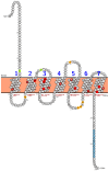
Numbering scheme for PMTRs. The most studied PMTR, AtPMTR1, was used as a reference to highlight topology and key conserved residues across 174 pPMTR sequences. Fully conserved residues are shown in red, mostly conserved in ECL and ICL domains in spermatophytes in orange, and cysteines involved in disulfide bridges in green. The most conserved residue for each TM domain, depicted as a squared box (light font color), was determined by consensus between our results and ConSurf [29]. These residues serve as the reference for the proposed PMTR numbering system based on Ballesteros and Weinstein [30]. Briefly, the first letter denotes the amino acid followed by its position in the protein, and then, in superscripts, the identifier starts with the TM number and ends with its position relative to the reference residue in that TM. That reference residue is arbitrarily assigned the number 50 [30]. The proposed reference residues are shown in red letters below each TM domain. YYSEM[KR]DAGFF is a motif completely conserved in seed plants (light blue). Domain predictions were derived from Tmbed [28] and visualized using PROTTER v1.0 [31].
Figure 5
Figure 5
Motifs discovered by MEME from the alignment of the 174 PMTR homolog sequences. A; Residues that interact with melatonin in site 1 and site 2 in AtPMTR1, CcPMTR1, and CcPMTR2 are represented with colors and shapes. Cysteine residues involved in disulfide bonds are highlighted. Residue coloring follows universalmotif package [32] defaults.
Figure 6
Figure 6
Putative docking sites for melatonin in PMTRs across Viridiplantae. Ligand poses obtained by AMDock [34] are presented for: M. commoda (McPMTR1), P. patens (PpPMTR1), M. polymorfa (MpPMTR1), S. moellendorffii (SmPMTR1), C. panzhihuaensis (CpPMTR1), A. thaliana (AtPMTR1, AtPMTR2), O. sativa (OsPMTR1), Z. mays (ZmPMTR1, ZmPMTR2), A. trichopoda (AtrPMTR1), M. esculenta (MePMTR1, MePMTR2), C. chinense (CcPMTR1, CcPMTR1), Cynara cardunculus (CcaPMTR1) D. carota (DcPMTR1), L. sativa (LsPMTR1), H. annus (HaPMTR1), and hMT1. Receptors are presented as cartoons where colors represent pLDDT scores for the predicted structure (dark blue: very high, blue: high, yellow: low, orange: very low). Ligand poses were classified based on binding affinity and colored accordingly as red (G7 kcal·mol−1), green (7<G6 kcal·mol−1), and pink (G>6 kcal·mol−1).
Figure 7
Figure 7
Binding pockets and ligand–receptor interactions for PMTRs of A. thaliana and C. chinense. (A), binding affinity distributions obtained from one-thousand Autodock Vina docking simulations for MT and receptors: AtPMTR1, CcPMTR1, and CcPMTR2. Site 1 (green) and site 2 (pink) for each receptor are shown, as well as the mean with its 95% confidence interval, boxplot, and histogram. A sample (n = 5) from the simulations was used to compare mean binding affinities between sites for each species independently. Distinct letters denote significant differences identified at a 95% confidence level using a Student’s t test. Receptor cartoon representations bound to melatonin in site 1 (green MT) and site 2 (pink MT) with ligand–receptor interactions for site 1 (superior green box, 1) and site 2 (inferior pink box, 2) for AtPMTR1 (B), CcPMTR1 (C), and CcPMTR2 (D). Hydrogen bonds (dark blue solid lines), π-π stacks (dashed green lines) and hydrophobic interactions (dashed grey lines) are shown. TM1-7, transmembrane domains; ECL2, extracellular loop 2. (E), residue relevance in the ligand–receptor interaction obtained through alanine-scanning and PLIP interaction profiler for AtPMTR1 (red), CcPMTR1 (green) and CcPMTR2 (blue). The horizontal red line is drawn at the 90th percentile.
Figure 8
Figure 8
Tunnel and ligand transport analysis for internal melatonin binding site 1 in A. thaliana and C. chinense. Receptors bound to melatonin were sliced longitudinally at the ligand plane, which exposes the internal cavities and tunnels: (A), AtPMTR1; (B), CcPMTR1; (C), CcPMTR2. Black boxes in the top-right corner display a lateral view of the cutting plane. Receptor residues (green) interacting with melatonin (pink) during its transit are represented as sticks. Tunnel radius and binding energy during melatonin trajectory through the tunnel for AtPMTR1, (D); CcPMTR1, (E); CcPMTR2, (F).
Figure 9
Figure 9
Protein–protein interaction networks for PMTRs in A. thaliana and C. chinense. (A), AtPMTR1; (B), CcPMTR1; (C), CcPMTR2.
Figure 10
Figure 10
A comparison between melatonin receptors in plants and animals. AtPMTR1 (left) and hMT1 (right) were used as representatives. Numbering sheme for residues is based on Ballesteros and Weinstein [30].

Similar articles

References

    1. Caspi Y., Pantazopoulou C.K., Prompers J.J., Pieterse C., Hulshoff Pol H., Kajala K. Why did glutamate, GABA, and melatonin become intercellular signalling molecules in plants? eLife. 2023;12:e83361. doi: 10.7554/eLife.83361. - DOI - PMC - PubMed
    1. Li D., Wei J., Peng Z., Ma W., Yang Q., Song Z., Sun W., Yang W., Yuan L., Xu X., et al. Daily rhythms of phytomelatonin signaling modulate diurnal stomatal closure via regulating reactive oxygen species dynamics in Arabidopsis. J. Pineal Res. 2020;68:e12640. doi: 10.1111/jpi.12640. - DOI - PubMed
    1. Wang L., Tanveer M., Wang H., Arnao M.B. Melatonin as a key regulator in seed germination under abiotic stress. J. Pineal Res. 2024;76:e12937. doi: 10.1111/jpi.12937. - DOI - PubMed
    1. Muhammad I., Ahmad S., Shen W. Melatonin-Mediated Molecular Responses in Plants: Enhancing Stress Tolerance and Mitigating Environmental Challenges in Cereal Crop Production. Int. J. Mol. Sci. 2024;25:4551. doi: 10.3390/ijms25084551. - DOI - PMC - PubMed
    1. Khan D., Cai N., Zhu W., Li L., Guan M., Pu X., Chen Q. The role of phytomelatonin receptor 1-mediated signaling in plant growth and stress response. Front. Plant Sci. 2023;14:1142753. doi: 10.3389/fpls.2023.1142753. - DOI - PMC - PubMed

LinkOut - more resources