. 2025 Jun 23;26(13):6003.
doi: 10.3390/ijms26136003.

Resolvin D2 and Its Effects on the Intestinal Mucosa of Crohn's Disease Patients: A Promising Immune Modulation Therapeutic Target

Affiliations

Resolvin D2 and Its Effects on the Intestinal Mucosa of Crohn's Disease Patients: A Promising Immune Modulation Therapeutic Target

Livia Bitencourt Pascoal et al. Int J Mol Sci. .

Abstract

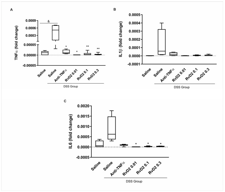
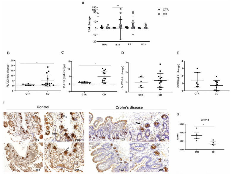
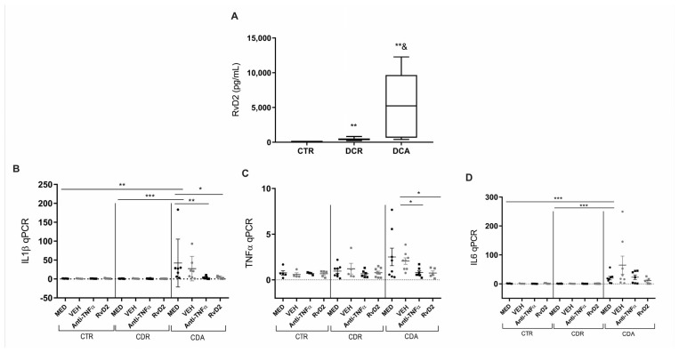
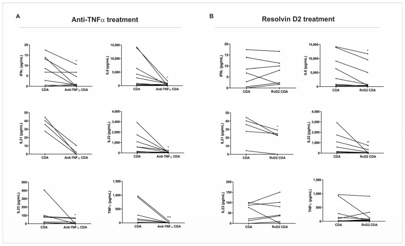
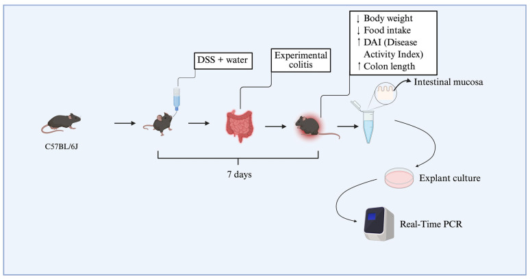
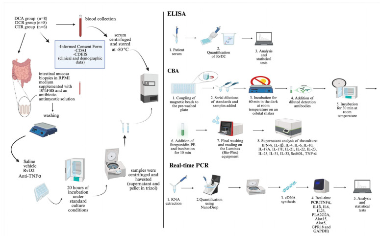
Crohn's disease (CD) is a chronic inflammatory disorder of the gastrointestinal tract that severely impacts patients' quality of life. Although current therapies have improved symptom management, they often fail to alter disease progression and are associated with immunosuppressive side effects. This study evaluated the immunomodulatory potential of resolvin D2 (RvD2), a pro-resolving lipid mediator, using a murine model of colitis and the ex vivo treatment of intestinal mucosal biopsies from CD patients, comparing its effects to those of conventional anti-TNFα therapy. To determine the optimal concentration of RvD2 for application in human tissue explant cultures, an initial in vitro assay was conducted using intestinal biopsies from mice with experimentally induced colitis. The explants were treated in vitro with varying concentrations of RvD2, and 0.1 μM emerged as an effective dose. This concentration significantly reduced the transcriptional levels of TNF-α (p = 0.004) and IL-6 (p = 0.026). Intestinal mucosal biopsies from fifteen patients with CD and seven control individuals were analyzed to validate RNA-sequencing data, which revealed dysregulation in the RvD2 biosynthetic and signaling pathways. The real-time PCR confirmed an increased expression of PLA2G7 (p = 0.02) and ALOX15 (p = 0.02), while the immunohistochemical analysis demonstrated the reduced expression of the RvD2 receptor GPR18 (p = 0.04) in intestinal tissues from CD patients. Subsequently, samples from eight patients with active Crohn's disease, eight patients in remission, and six healthy controls were used for the serum analysis of RvD2 by ELISA, in vitro treatment of intestinal biopsies with RvD2 or anti-TNF, followed by transcriptional analysis, and a multiplex assay of the explant culture supernatants. The serum analysis demonstrated elevated RvD2 levels in CD patients both with active disease (p = 0.02) and in remission (p = 0.002) compared to healthy controls. The ex vivo treatment of intestinal biopsies with RvD2 decreased IL1β (p = 0.04) and TNFα (p = 0.02) transcriptional levels, comparable to anti-TNFα therapy. Additionally, multiplex cytokine profiling confirmed a reduction in pro-inflammatory cytokines, including IL-6 (p = 0.01), IL-21 (p = 0.04), and IL-22 (p = 0.009), in the supernatant of samples treated with RvD2. Altogether, these findings suggest that RvD2 promotes the resolution of inflammation in CD and supports its potential as a promising therapeutic strategy.

Keywords: Crohn’s disease; immunoregulation; pro-resolving lipid mediator; resolvin D2.


Conflict of interest statement

The authors declare no conflicts of interest.

Figures

Figure 1
Figure 1
Inflammatory mediators in the intestinal mucosa of DSS-induced colitis after treatment with RvD2 and anti-TNFα in explant culture. mRNA levels (qPCR) of TNFα (A), IL1β (B), and IL6 (C) were measured in the intestinal mucosa of mice from the DSS group compared to the DSS-non-treated group. DSS = dextran sulfate sodium. Saline = biopsies treated with saline. Anti-TNFα = biopsies treated with infliximab at a 100 µg/mL concentration. RvD2 0.01 = biopsies treated with resolvin D2 at a concentration of 0.01 µM. RvD2 0.1 = biopsies treated with resolvin D2 at a concentration of 0.1 µM. RvD2 0.3 = biopsies treated with resolvin D2 at a concentration of 0.3 µM. n = 6. Data are presented as the mean ± SEM. & p < 0.05 was considered statistically significant compared to the DSS-non-treated group. * p < 0.05 and ** p < 0.01 were considered statistically significant compared to the saline-treated DSS group.
Figure 2
Figure 2
Inflammatory mediators and RvD2 biosynthesis pathway in the ileal mucosa of Crohn’s disease patients. (A) Transcriptional analysis of pro-inflammatory cytokines. TNFα, IL1β, IL6, and L23 mRNA levels (fold change) were measured in the ileal mucosa of Crohn’s disease patients (CD) compared to controls (CTR). (BE) Transcriptional analysis of genes involved in the RvD2 biosynthesis pathway: (B) PLA2G, (C) 15-LOX, (D) 5-LOX, and (E) GPR18 mRNA levels (fold change) in the ileal mucosa of Crohn’s disease patients (CD) compared to controls (CTR). (F) Immunohistochemical analysis of GPR18 was performed on paraffin-embedded slides from the ileal mucosa of both Crohn’s disease and control groups. The white arrows indicate positive epithelial cells, and the black arrows signal positive cells from the lamina propria; 10× and 20× objective lenses were used as indicated in each image. (G) Quantitative analysis of RvD2 staining in the ileal mucosa of Crohn´s disease patients (CD) compared to controls (CTR). Results are presented as the mean ± SEM. CTR = control group; CD = Crohn’s disease group. n = 7 for CTR and n = 15 for CD in the transcriptional analyses; n = 4 for CTR and n = 4 for CD in the immunohistochemical analysis. * p < 0.05 and ** p < 0.01 were considered statistically significant compared to the CTR group.
Figure 3
Figure 3
Serum resolvin D2 levels and transcriptional analysis of pro-inflammatory cytokines in Crohn’s Disease: Effects of in vitro treatment with resolvin D2 or anti-TNF therapy. (A) Crohn’s disease patients with active disease (CDA group) and those in remission (CDR group) exhibited increased serum levels of resolvin D2 compared to control subjects (CTR). ** p < 0.01 vs. CTR; p = 0.02 for CDA vs. CDR. (BD) Transcriptional analysis of pro-inflammatory cytokines in Crohn’s disease following in vitro treatment with resolvin D2 or anti-TNF as an immunomodulatory agent: (B) IL1β transcriptional levels were significantly increased in the MED-treated CDA group compared to the MED CTR and MED CDR groups. Both anti-TNF and resolvin D2 treatments were effective in reducing IL1β expression compared to the MED CDA group; (C) TNFα transcriptional levels were significantly decreased in the treatment with anti-TNF or resolvin D2 conditions compared to VEH-treated CDA condition; (D) IL6 transcriptional levels were significantly increased in the MED CDA group compared to MED CTR and MED CDR groups; however, neither anti-TNF nor resolvin D2 treatment modulated IL6 expression compared to the MED CDA condition. CDA = Crohn’s disease in active disease. CDR = Crohn’s disease in remission. CTR = Control. MED = medium. VEH = vehicle. Anti-TNFα = infliximab. RvD2 = resolvin D2. CDA n = 8. CDR n = 8. CTR n = 6. * p < 0.05, ** p < 0.01 and *** p < 0.001 were considered statistically significant compared to the CTR group. & p < 0.05 were considered statistically significant compared to the CDR group.
Figure 4
Figure 4
Protein expression of pro-inflammatory cytokines in supernatants of intestinal biopsy culture from active Crohn’s disease patients. Intestinal biopsies from Crohn’s disease patients in active disease (CDA group) were treated with resolving D2 (RvD2) or anti-TNFα (infliximab), and supernatants were collected for cytokine quantification using a multiplex assay. (A) Treatment with anti-TNFα significantly reduced the levels of IFN-γ, IL-6, IL-22, IL-23, and TNF-α compared to the respective untreated CDA group. (B) Treatment with resolvin D2 significantly decreased IL-6, IL-21, and IL-22 levels compared to the respective untreated CDA group. CDA = Crohn’s disease in active disease. Anti-TNFα CDA = Supernatant of intestinal biopsies culture from CDA patients treated with infliximab. RvD2 CDA = Supernatant of intestinal biopsies culture from CDA patients treated with resolvin D2. CDA n = 8. Anti-TNFα CDR n = 8. RvD2 CDA n = 8. * p < 0.05, ** p < 0.01 and *** p < 0.001 were considered statistically significant.
Figure 5
Figure 5
Schematic representation of the dose-response experimental design. C57BL/6J mice were divided into two groups, receiving or not receiving DSS diluted in drinking water for seven days. This resulted in weight loss, reduced food intake, and an increase in the disease activity index (DAI). Colon shortening was confirmed at euthanasia. Subsequently, colons were collected and cultured as explants, and three concentrations of resolvin D2 (0.01 μM, 0.1 μM, and 0.3 μM) were tested. The diagram illustrates the main stages of the protocol, including induction of experimental colitis with DSS, validation of methodological efficacy, tissue sample collection for explant culture, and subsequent gene expression analysis by real-time PCR. ↑: increase. ↓ decrease.
Figure 6
Figure 6
Schematic representation of the ex vivo treatment of the intestinal mucosa biopsies with resolvin D2 or anti-TNFα, and the processing of the samples. ELISA and CBA assays were employed for protein analysis, and real-time PCR was performed for transcriptional analysis.

Similar articles

References

    1. Carroll D., Kavalukas S. Management of complications in Crohn’s disease. Adv. Surg. 2024;58:19–34. doi: 10.1016/j.yasu.2024.04.002. - DOI - PubMed
    1. Petagna L., Antonelli A., Ganini C., Bellato V., Campanelli M., Divizia A., Efrati C., Franceschilli M., Guida A.M., Ingallinella S., et al. Pathophysiology of Crohn’s disease inflammation and recurrence. Biol. Direct. 2020;15:23. doi: 10.1186/s13062-020-00280-5. - DOI - PMC - PubMed
    1. Ng S.C., Shi H.Y., Hamidi N., Underwood F.E., Tang W., Benchimol E.I., Panaccione R., Ghosh S., Wu J.C.Y., Chan F.K.L., et al. Worldwide incidence and prevalence of inflammatory bowel disease in the 21st century: A systematic review of population-based studies. Lancet. 2018;390:2769–2778. doi: 10.1016/S0140-6736(17)32448-0. - DOI - PubMed
    1. Veauthier B., Hornecker J.R. Crohn’s disease: Diagnosis and management. Am. Fam. Phys. 2018;98:661–669. - PubMed
    1. Shah S.C., Itzkowitz S.H. Colorectal cancer in inflammatory bowel disease: Mechanisms and management. Gastroenterology. 2022;162:715–730.e3. doi: 10.1053/j.gastro.2021.10.035. - DOI - PMC - PubMed

MeSH terms

LinkOut - more resources