. 2025 Jun 24;26(13):6049.
doi: 10.3390/ijms26136049.

Type I Interferons in SARS-CoV-2 Cutaneous Infection: Is There a Role in Antiviral Defense?

Affiliations

Type I Interferons in SARS-CoV-2 Cutaneous Infection: Is There a Role in Antiviral Defense?

Tatiana Mina Yendo et al. Int J Mol Sci. .

Abstract

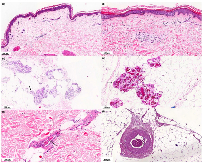
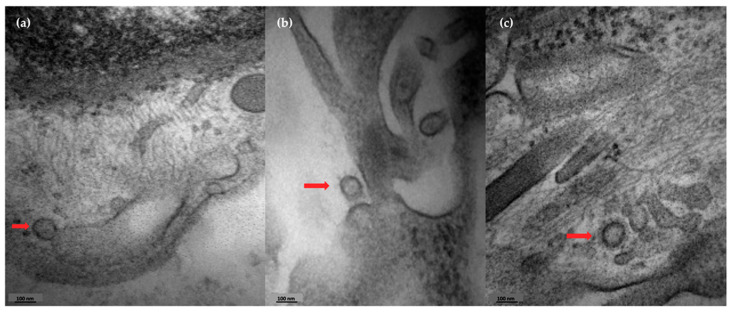
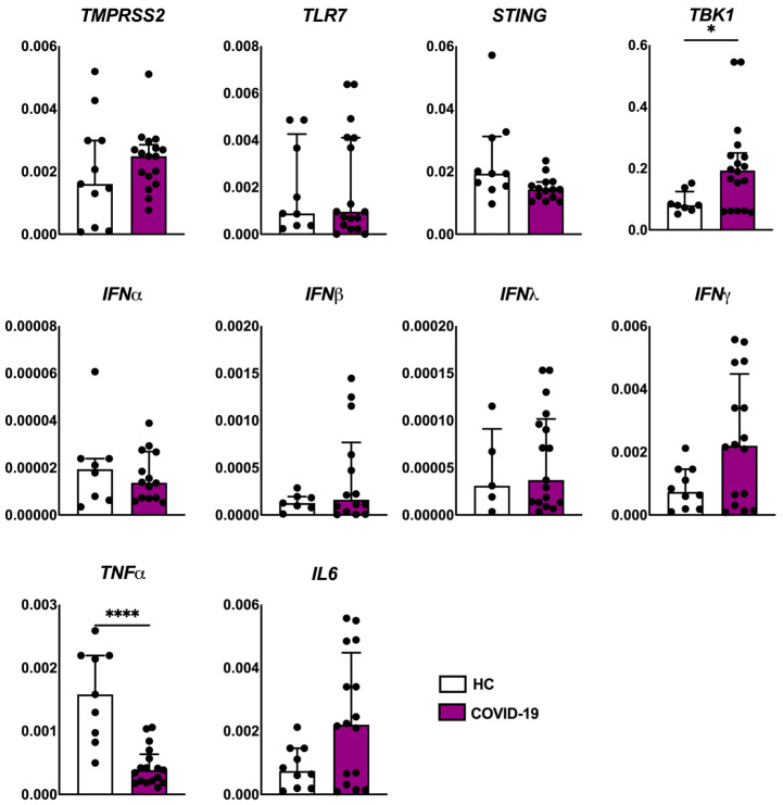
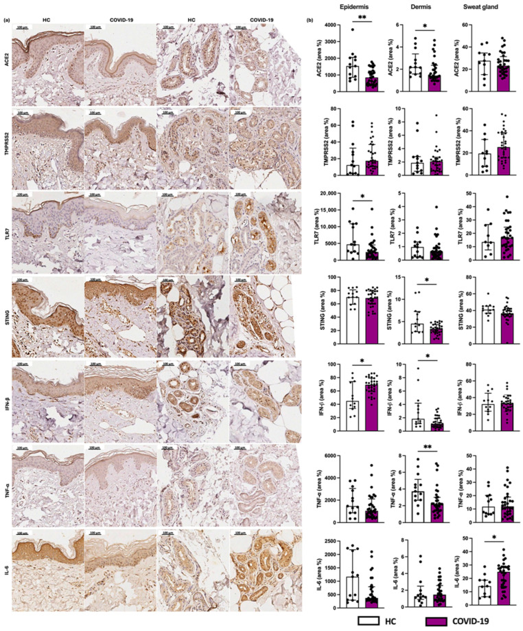
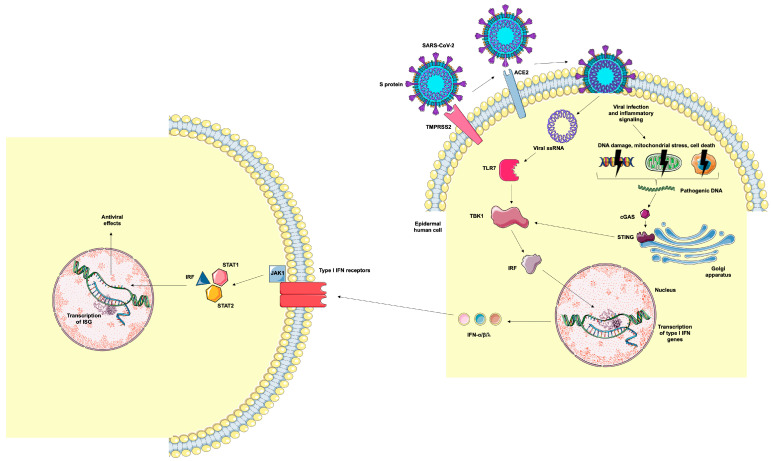
SARS-CoV-2, a β-coronavirus, primarily affects the lungs, with non-specific lesions and no cytopathic viral effect in the skin. Cutaneous antiviral mechanisms include activation of TLR/IRF pathways and production of type I IFN. We evaluated the antiviral mechanisms involved in the skin of COVID-19 patients, including skin samples from 35 deceased patients who had contracted COVID-19 before the launch of the vaccine. Detection of SARS-CoV-2 in the skin was performed using transmission electron microscopy and RT-qPCR. Microscopic and molecular effects of the virus in skin were evaluated by histopathology, RT-qPCR, and immunohistochemistry (IHC). The results revealed the presence of SARS-CoV-2 and microscopic changes, including microvascular hyaline thrombi, perivascular dermatitis, and eccrine gland necrosis. There was increased transcription of TBK1 and a reduction in transcription of TNFα by RT-qPCR in the COVID-19 group. IHC revealed reduced expression of ACE2, TLR7, and IL-6, and elevated expression of IFN-β by epidermal cells. In the dermis, there was decreased expression of STING, IFN-β, and TNF-α and increased expression of IL-6 in sweat glands. Our results highlight the role of type I IFN in the skin of COVID-19 patients, which may modulate the cutaneous response to SARS-CoV-2.

Keywords: COVID-19; SARS-CoV-2; STING; skin.


Conflict of interest statement

The authors declare no conflicts of interest.

Figures

Figure 1
Figure 1
Histopathological analysis of the skin of the COVID-19 group. Photographs of skin specimens from healthy controls (HCs) and COVID-19 patients by hematoxylin and eosin staining. (a) Absence of significant histological changes (×100). (b) Mild to moderate perivascular dermatitis (×100). (c) Vacuolar degeneration of sweat glands cells (black arrows) (×100). (d) Necrosis of eccrine glands associated with hyaline thrombi (black arrows) in the peri glandular vessels (×100). (e) Hyaline thrombus in dermal vessel (black arrow) (×100). (f) Hyaline thrombus in a larger vessel in the hypodermis (black arrow) (×200).
Figure 2
Figure 2
Skin detection of coronavirus by transmission electron microscopy. Photographs of the presence of coronavirus in skin sections from a COVID-19 patient. (a) Presence of viral budding (red arrow). (b,c) Viral particles detaching from cellular structures (red arrows).
Figure 3
Figure 3
Evaluation of the expression of viral receptor (TMPRSS2 and TLR7), antiviral markers (STING, TBK1, IFNα, IFNβ, IFNλ and IFNγ), and pro-inflammatory markers (TNFα and IL6) genes in the skin of patients with COVID-19 (n = 18) compared to healthy controls (HCs, n = 10). Values are expressed as median with interquartile range. * p < 0.05; **** p < 0.0001.
Figure 4
Figure 4
Evaluation of the expression of viral receptors. (a) Photographs of skin specimens from healthy controls (HCs) and COVID-19 patients: ACE2, TMPRSS2, and TLR7 (×200). (b) Expression of ACE2, TMPRSS2, and TLR7 in the epidermis (area %), dermis (area %), and sweat glands (area %) in COVID-19 patients (n = 32) in comparison to the HC group (n = 13). Values are expressed in median with interquartile range. * p < 0.05; ** p < 0.01.
Figure 5
Figure 5
Representation of epidermal human cell infection by SARS-CoV-2 and antiviral mechanisms. TMPRSS2 cleaves the viral S protein into S1 and S2 subunits. The S1 protein binds to ACE2 and S2 promotes the fusion of viral and cellular membranes, allowing the internalization of the virus. Viral RNA binds to TLR7, resulting in the activation of TBK1 and IRF, which are translocated to the cell nucleus, resulting in the gene transcription of type I IFN (α, β, and λ). Cellular infection by the virus and the inflammatory milieu can lead to DNA damage, mitochondrial stress, and cell death, resulting in the generation of cytosolic DNA. These pathogenic DNAs bind to cGAS, which activates the STING signaling pathway, resulting in the transcription of type I IFN genes. Type I IFNs bind to cellular receptors, activating the JAK-STAT intracellular pathway that results in the translocation of IRF to the nucleus and the transcription of ISGs. Image provided by Servier Medical Art (https://smart.servier.com/), accessed on 20 May 2025, licensed under CC BY 4.0 (https://creativecommons.org/licenses/by/4.0/), accessed on 20 May 2025.

Similar articles

References

    1. Hu B., Guo H., Zhou P., Shi Z.L. Characteristics of SARS-CoV-2 and COVID-19. Nat. Rev. Microbiol. 2021;19:141–154. doi: 10.1038/s41579-020-00459-7. - DOI - PMC - PubMed
    1. Yang H., Rao Z. Structural biology of SARS-CoV-2 and implications for therapeutic development. Nat. Rev. Microbiol. 2021;19:685–700. doi: 10.1038/s41579-021-00630-8. - DOI - PMC - PubMed
    1. Beyerstedt S., Casaro E.B., Rangel É. COVID-19: Angiotensin-converting enzyme 2 (ACE2) expression and tissue susceptibility to SARS-CoV-2 infection. Eur. J. Clin. Microbiol. Infect. Dis. 2021;40:905–919. doi: 10.1007/s10096-020-04138-6. - DOI - PMC - PubMed
    1. Zhou Z., Ren L., Zhang L., Zhong J., Xiao Y., Jia Z., Guo L., Yang J., Wang C., Jiang S., et al. Heightened Innate Immune Responses in the Respiratory Tract of COVID-19 Patients. Cell Host Microbe. 2020;27:883–890.E2. doi: 10.1016/j.chom.2020.04.017. - DOI - PMC - PubMed
    1. Lipsker D. Paraviral eruptions in the era of COVID-19: Do some skin manifestations point to a natural resistance to SARS-CoV-2? Clin. Dermatol. 2020;38:757–761. doi: 10.1016/j.clindermatol.2020.06.005. - DOI - PMC - PubMed

MeSH terms