. 2025 Jun 24;26(13):6067.
doi: 10.3390/ijms26136067.

Naturally Occurring Angiotensin Peptides Enhance the SARS-CoV-2 Spike Protein Binding to Its Receptors

Affiliations

Naturally Occurring Angiotensin Peptides Enhance the SARS-CoV-2 Spike Protein Binding to Its Receptors

Katelin X Oliveira et al. Int J Mol Sci. .

Abstract

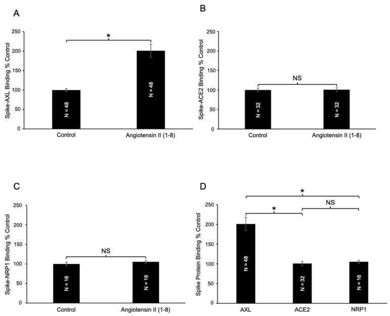
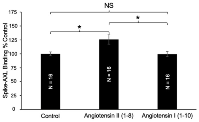
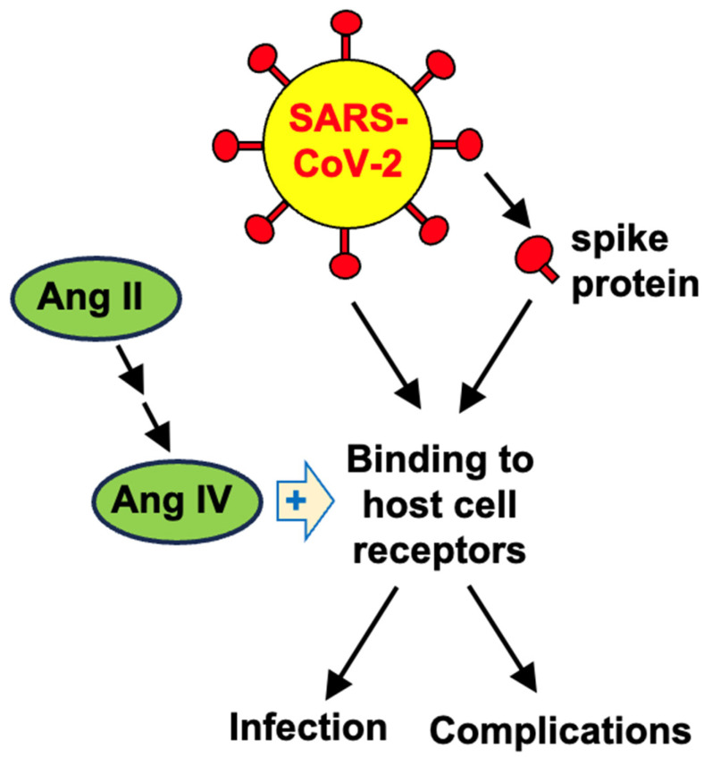
Severe Acute Respiratory Syndrome Coronavirus 2 (SARS-CoV-2), the virus responsible for Coronavirus Disease 2019 (COVID-19), utilizes its spike protein to infect host cells. In addition to angiotensin-converting enzyme 2 (ACE2) and neuropilin-1 (NRP1), AXL acts as a spike protein receptor and mediates infection, especially in respiratory cells with low ACE2 expression. Angiotensin II (1-8) can be cleaved into shorter peptides within the biological system. Antibody-based binding assays showed that angiotensin II causes a two-fold increase in the binding between the spike protein and AXL, but not ACE2 or NRP1. While a longer peptide, angiotensin I (1-10), did not affect the spike-AXL binding, shorter lengths of angiotensin peptides exhibited enhancing effects. The C-terminal deletions of angiotensin II to angiotensin (1-7) or angiotensin (1-6) resulted in peptides with enhanced activity toward spike-AXL binding with a similar capacity as angiotensin II. In contrast, the N-terminal deletions of angiotensin II to angiotensin III (2-8) or angiotensin IV (3-8) as well as the N-terminal deletions of angiotensin (1-7) to angiotensin (2-7) or angiotensin (5-7) produced peptides with a more potent ability to enhance spike-AXL binding (2.7-fold increase with angiotensin IV). When valine was substituted for tyrosine at position 4 in angiotensin II or when tyrosine at position 4 was phosphorylated, spike-AXL binding was increased, suggesting that modifications to tyrosine trigger enhancement. Angiotensin IV also enhances spike protein binding to ACE2 and NRP1. Thus, angiotensin peptides may contribute to COVID-19 pathogenesis by enhancing spike protein binding and thus serve as therapeutic targets.

Keywords: AXL; COVID-19; SARS-CoV-2; angiotensin; peptide; spike protein.


Conflict of interest statement

The authors declare no conflicts of interest. The funders had no role in the design of the study; in the collection, analyses, or interpretation of data; in the writing of the manuscript; or in the decision to publish the results.

Figures

Figure 1
Figure 1
Structures of the angiotensin peptides. This figure illustrates the structures of the naturally occurring angiotensin peptides relevant to the experiments performed in this study. It also outlines how angiotensin I (1–10), angiotensin II (1–8), angiotensin (1–7), angiotensin III (2–8), and angiotensin IV (3–8) are physiologically synthesized in the biological system.
Figure 2
Figure 2
Effects of angiotensin II (1–8) on spike protein binding. (A) The addition of angiotensin II (1–8) significantly enhances spike–AXL binding. In contrast, angiotensin II (1–8) did not significantly alter (B) spike–ACE2 binding or (C) spike–NRP1 binding, respectively. (D) The effect of angiotensin II (1–8) on spike protein binding across three different receptors. Angiotensin II (1–8) selectively enhanced spike–AXL binding and did not influence spike–ACE2 binding or spike–NRP1 binding. The bar graphs represent means ± SEM. An asterisk (*) denotes that the values are significantly different from each other at p < 0.05, and “NS” indicates “not significant”.
Figure 3
Figure 3
Effects of angiotensin I (1–10) on spike–AXL binding. Only angiotensin II (1–8) increases spike–AXL binding, while angiotensin I (1–10) does not. The bar graphs represent means ± SEM. An asterisk (*) denotes that the values are significantly different from each other at p < 0.05, and “NS” indicates “not significant”.
Figure 4
Figure 4
Effects of angiotensin (1–7) and angiotensin (1–6) on spike–AXL binding. Angiotensin II (1–8), angiotensin (1–7), and angiotensin (1–6) all significantly enhance spike–AXL binding. However, when compared to each other, the enhancing effects of angiotensin II (1–8), angiotensin (1–7), and angiotensin (1–6) are not significantly different from one another. The bar graphs represent means ± SEM. An asterisk (*) denotes that the values are significantly different from each other at p < 0.05, and “NS” indicates “not significant”.
Figure 5
Figure 5
Effects of angiotensin III (2–8) and angiotensin IV (3–8) on spike–AXL Binding. (A) Both angiotensin II (1–8) and angiotensin III (2–8) enhance spike–AXL binding, with angiotensin III (2–8) enhancing spike–AXL binding more potently than angiotensin II (1–8). (B) Both angiotensin II (1–8) and angiotensin IV (3–8) significantly increase spike–AXL binding, with angiotensin IV (3–8) enhancing spike–AXL binding more potently than angiotensin II (1–8). (C) Angiotensin IV (3–8) enhances spike–AXL binding more potently than angiotensin III (2–8). The bar graphs represent means ± SEM. An asterisk (*) denotes that the values are significantly different from each other at p < 0.05.
Figure 6
Figure 6
Effects of angiotensin (2–7) and angiotensin (5–7) on spike–AXL binding. Angiotensin II (1–8), angiotensin (2–7), and angiotensin (5–7) all significantly enhance the spike–AXL binding. The enhancing effects of angiotensin (2–7) and angiotensin (5–7) are more potent than those of angiotensin II (1–8), and the effects of angiotensin (2–7) and angiotensin (5–7) are comparable to each other. The bar graphs represent means ± SEM. An asterisk (*) denotes that the values are significantly different from each other at p < 0.05, and “NS” indicates “not significant”.
Figure 7
Figure 7
Effects of angiotensin II (1–8) analogs on Spike–AXL binding. Angiotensin II (1–8) and its analogs, Sar1, Ala8 angiotensin II (1–8), Val4 angiotensin II (1–8), and Tyr(PO3H2)4 angiotensin II (1–8), all enhance the spike–AXL binding. The enhancing effects of the analogs on spike–AXL binding were significantly greater than those of angiotensin II (1–8), but the differences among the effects of the analogs were not significant. The bar graphs represent means ± SEM. An asterisk (*) denotes that the values are significantly different from each other at p < 0.05, and “NS” indicates “not significant”.
Figure 8
Figure 8
Effects of angiotensin IV (3–8) on spike–AXL binding to host cell receptors. (A) Angiotensin IV (3–8) significantly enhanced spike–AXL binding. (B) Angiotensin IV (3–8) significantly enhanced spike–ACE2 binding. (C) Angiotensin IV (3–8) significantly enhanced spike–NRP1 binding. (D) Although angiotensin IV (3–8) enhanced spike–AXL binding, spike–ACE2 binding, and spike–NRP1 binding, this peptide most potently enhanced spike–AXL binding. The bar graphs represent means ± SEM. An asterisk (*) denotes that the values are significantly different from each other at p < 0.05, and “NS” indicates “not significant”.
Figure 9
Figure 9
A scheme summarizing the key results of the present study.
Figure 10
Figure 10
Principles of the spike protein–AXL binding assay. The RayBio COVID-19 Spike-AXL Binding Assay Kit contains AXL protein bound to the bottom of the microplate wells. SARS-CoV-2 spike protein S1 is added, washed, and the bound spike protein S1 to AXL is detected using anti-spike protein S1 IgG antibody and HRP-conjugated anti-IgG. The addition of the TMB substrate results in HRP catalyzing the reaction to produce a detectable color using an absorbance microplate reader. Created in BioRender. Oliveira, K. (2025) https://BioRender.com/s0zewuu.

Update of

Similar articles

Cited by

References

    1. Jackson C.B., Farzan M., Chen B., Choe H. Mechanisms of SARS-CoV-2 entry into cells. Nat. Rev. Mol. Cell Biol. 2022;23:3–20. doi: 10.1038/s41580-021-00418-x. - DOI - PMC - PubMed
    1. Lamers M.M., Haagmans B.L. SARS-CoV-2 pathogenesis. Nat. Rev. Microbiol. 2022;20:270–284. doi: 10.1038/s41579-022-00713-0. - DOI - PubMed
    1. Forrester S.J., Booz G.W., Sigmund C.D., Coffman T.M., Kawai T., Rizzo V., Scalia R., Eguchi S. Angiotensin II signal transduction: An update on mechanisms of physiology and pathophysiology. Physiol. Rev. 2018;98:1627–1738. doi: 10.1152/physrev.00038.2017. - DOI - PMC - PubMed
    1. Paz Ocaranza M., Riquelme J.A., García L., Jalil J.E., Chiong M., Santos R.A.S., Lavandero S. Counter-regulatory renin-angiotensin system in cardiovascular disease. Nat. Rev. Cardiol. 2020;17:116–129. doi: 10.1038/s41569-019-0244-8. - DOI - PMC - PubMed
    1. Benigni A., Cassis P., Remuzzi G. Angiotensin II revisited: New roles in inflammation, immunology and aging. EMBO Mol. Med. 2010;2:247–257. doi: 10.1002/emmm.201000080. - DOI - PMC - PubMed

MeSH terms

LinkOut - more resources