. 2025 Jun 27;26(13):6188.
doi: 10.3390/ijms26136188.

The Production of Recombinant Azurin from Pseudomonas aeruginosa and Its Ability to Induce Apoptosis in Various Breast Cancer Cell Lines

Affiliations

The Production of Recombinant Azurin from Pseudomonas aeruginosa and Its Ability to Induce Apoptosis in Various Breast Cancer Cell Lines

Tomasz Kowalczyk et al. Int J Mol Sci. .

Abstract

Azurin is a copper-containing redox protein naturally produced by Pseudomonas aeruginosa, which has shown promising activity against human cancer cells by inducing apoptosis. The present study describes the design of a recombinant vector, pT7-MAT-Tag-2-Azu, for azurin production in E. coli cells. The cytotoxic effects of purified azurin were tested on three breast cancer cell lines (MCF-7, MDA-MB-231, and HCC38) and a normal breast epithelial cell line (MCF10A) using the MTT assay. The results showed cytotoxicity against cancer cell lines with minimal effects on normal cells. Further analysis showed that azurin induced apoptosis through mitochondrial pathways, as evidenced by increased expression of apoptosis-related genes (Bax, TP53, Apaf-1, caspase-3, -8, -9) and their corresponding proteins, elevated levels of reactive oxygen species (ROS), and DNA damage, mitochondrial membrane potential (MMP), or brine shrimp lethality assay. Furthermore, in silico molecular docking, simulations predicted a stable, electrostatically driven interaction between azurin and the p53 protein, providing a structural basis for its mechanism of action. These findings suggest that recombinant azurin may serve as a potential therapeutic agent for breast cancer after further multifaceted research.

Keywords: anticancer; apoptosis; azurin; breast cancer; recombinant proteins.


Conflict of interest statement

The authors declare that they have no known competing financial interests or personal relationships that could have appeared to influence the work reported in this paper.

Figures

Figure 1
Figure 1
Recombinant vector pT7-MAT-Tag-2-Azu (SnapGene® software, www.snapgene.com, ver. 4.2.11).
Figure 2
Figure 2
Structural model of the Azurin-p53 complex predicted by HADDOCK. The top-scoring model from the best-ranked cluster is shown in a cartoon representation. Azurin (orange) is docked into a surface groove of the p53 DNA-binding domain (cyan). The catalytic copper ion (Cu2⁺) in azurin is shown as a yellow sphere, and the structural zinc ion (Zn2⁺) in p53 is shown as a magenta sphere. The image was generated using PyMOL (version 3.1).
Figure 3
Figure 3
Result of analysis of the pT7-MAT-Tag-2-Azu recombinant vector construct. (A) Hydrolysis by restriction enzymes, M-DNA size marker, 1–5—results of hydrolysis of independent recombinants; (B) PCR analysis, M-DNA size marker, 1–2—PCR on recombinant vectors, 3—negative control without vector.
Figure 4
Figure 4
(A) SDS-PAGE of protein isolated from transformed E. coli, M-Protein ladder, 1—proteins isolated from non-IPTG-induced bacteria, 2—proteins isolated from IPTG-induced bacteria, 3—flow through (IMAC purification), 4—purified azurin; (B) Western blot analysis of affinity chromatography purified recombinant azurin, M—prestained protein marker, 1–2—purified recombinant azurin (independent cell lysates), 3—negative control (isolated proteins from untransformed E. coli cells).
Figure 5
Figure 5
Viability of the normal MCF10A cell line (A), and the breast cancer cell lines HCC38 (B), MDA-MB-231 (C), and MCF-7 (D) after treatment with recombinant azurin. Cells were treated with various concentrations of recombinant protein. The cytotoxic effect was tested after 24 h of exposure. Data shown are the mean values of three independent experiments. ** p < 0.01, *** p < 0.001.
Figure 6
Figure 6
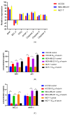
Effect of recombinant azurin on the expression of Bax, Bcl-2, TP53, Apaf-1, caspase-3, caspase-8, and caspase-9 in MCF-7, MDA-MB-231, and HCC38 cancer cells. Expression at the mRNA (A) and protein levels (B,C). Results are mean ± SD from three independent experiments. Protein levels of BAX, BCL-2, p53, and APAF-1 in HCC38, MDA-MB-231, and MCF-7 cancer cell lines determined by ELISA assay after treatment with recombinant azurin protein (IC50 for 24 h). Results are mean ± SD from four independent experiments. * p < 0.05, ** p < 0.001, *** p < 0.001.
Figure 7
Figure 7
(A) Comet assay showing recombinant protein azurin-induced DNA breaks in MCF-7, MDA-MB-231, and HCC38 cells for 24 h. (B) In untreated MCF-7, MDA-MB-231, and HCC38 cells, a small amount of inherent endogenous DNA damage could be seen. Results are given as mean ± SD from three independent experiments. ** p < 0.01, *** p < 0.001.
Figure 8
Figure 8
ROS levels of MCF-7, MDA-MB-231, and HCC38 cancer cells treated with the IC50 of recombinant azurin protein for 2, 8, 12, and 24 h along with controls were measured after staining with 2′,7′—Dichlorofluorescin diacetate (DCFH-DA). Data are given as the mean of three independent experiments. * p < 0.05, ** p < 0.01.
Figure 9
Figure 9
Caspase 3/7 activity (RFU) in MCF-7, MDA-MB-231, and HCC38 cancer cells treated with IC50 concentration of recombinant azurin protein for 24 h. The activities of caspase 3/7 in treated and untreated cells were then determined by luminescence assay. Data shown are the mean of three independent experiments. ** p < 0.01, *** p < 0.001.
Figure 10
Figure 10
Effect of recombinant azurin on the mitochondrial membrane potential (ΔΨm) in breast cancer cells. MCF-7, MDA-MB-231, and HCC38 cells were treated with azurin at their respective IC50 concentrations for 24 h. The mitochondrial membrane potential was quantified using the JC-1 probe. Data are expressed as the mean ratio of red (J-aggregate) to green (JC-1 monomer) fluorescence ± SD from three independent experiments. Asterisks indicate a statistically significant decrease compared to the corresponding untreated control group (*** p < 0.001).
Figure 11
Figure 11
General toxicity of recombinant azurin evaluated by the brine shrimp (Artemia salina) lethality assay. Nauplii were exposed to a range of azurin concentrations for 24 h. The columns represent the mean percentage of mortality ± standard deviation (SD) from three independent experiments. A vehicle control (protein buffer) and a positive control (1 mg/mL potassium dichromate, K2Cr2O7) were included to validate the assay. Statistical significance was determined by comparing each azurin treatment group to the vehicle control group using a one-way ANOVA followed by Dunnett’s post hoc test. (* p < 0.05; ** p < 0.01; *** p < 0.001).

Similar articles

References

    1. Akbarian M. Insulin therapy; a valuable legacy and its future perspective. Int. J. Biol. Macromol. 2021;181:1224–1230. doi: 10.1016/j.ijbiomac.2021.05.052. - DOI - PubMed
    1. Maiorella B., Inlow D., Shauger A., Harano D. Large-Scale Insect Cell-Culture for Recombinant Protein Production. Bio/Technol. 1988;6:1406–1410. doi: 10.1038/nbt1288-1406. - DOI
    1. Hacker D.L., Ordóñez-Morán P. Large-Scale Production of Recombinant Noggin and R-Spondin1 Proteins Required for the Maintenance of Stem Cells in Intestinal Organoid Cultures. Methods Mol. Biol. 2020;2171:171–184. doi: 10.1007/978-1-0716-0747-3_10. - DOI - PubMed
    1. Diamos A.G., Hunter J.G.L., Pardhe M.D., Rosenthal S.H., Sun H., Foster B.C., DiPalma M.P., Chen Q., Mason H.S. High Level Production of Monoclonal Antibodies Using an Optimized Plant Expression System. Front. Bioeng. Biotechnol. 2020;7:504837. doi: 10.3389/fbioe.2019.00472. - DOI - PMC - PubMed
    1. Camire R.M. Blood coagulation factor X: Molecular biology, inherited disease, and engineered therapeutics. J. Thromb. Thrombolysis. 2021;52:383–390. doi: 10.1007/s11239-021-02456-w. - DOI - PMC - PubMed

MeSH terms