. 2025 Jun 27;26(13):6232.
doi: 10.3390/ijms26136232.

Multispectral Pulsed Photobiomodulation Enhances Diabetic Wound Healing via Focal Adhesion-Mediated Cell Migration and Extracellular Matrix Remodeling

Affiliations

Multispectral Pulsed Photobiomodulation Enhances Diabetic Wound Healing via Focal Adhesion-Mediated Cell Migration and Extracellular Matrix Remodeling

Jihye Choi et al. Int J Mol Sci. .

Abstract

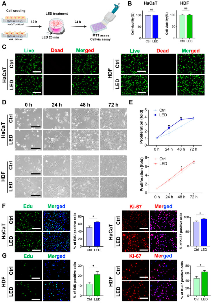
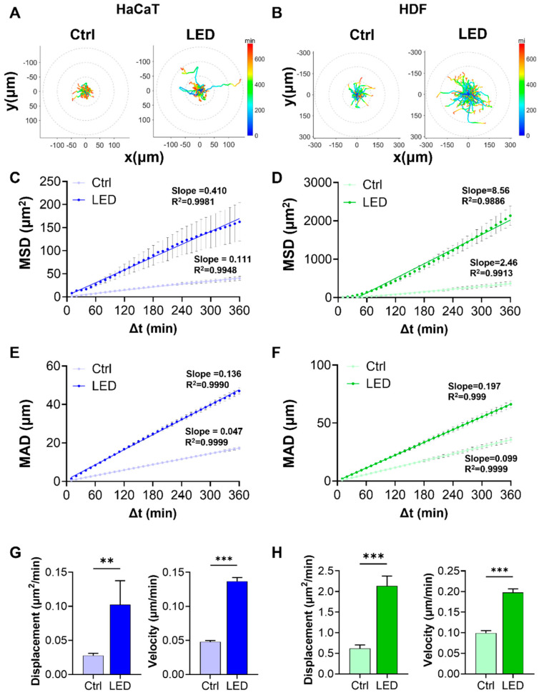
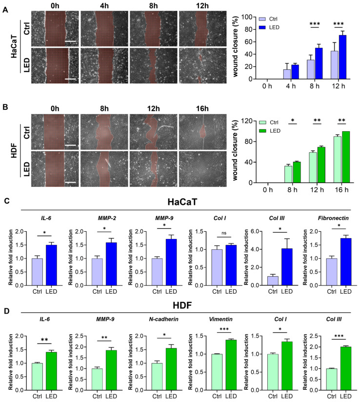
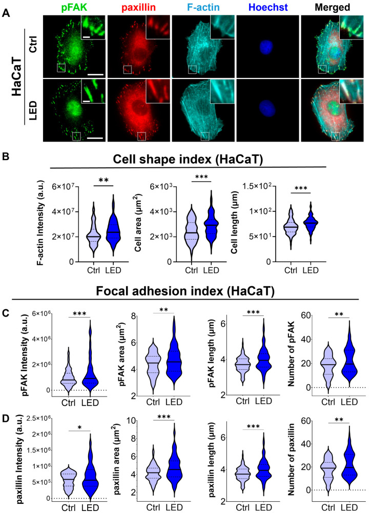
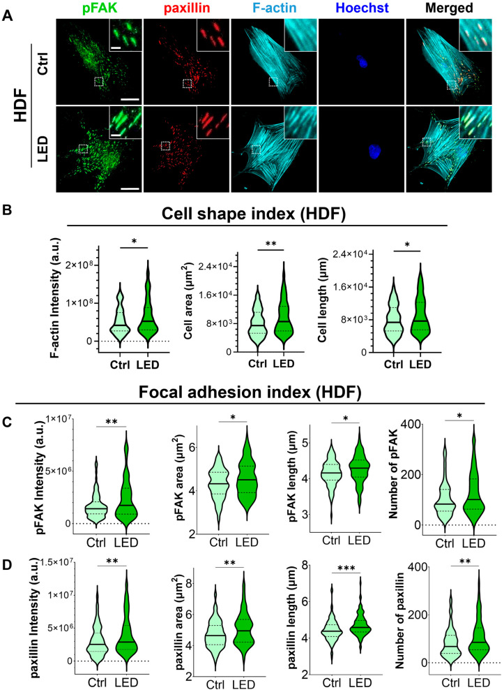
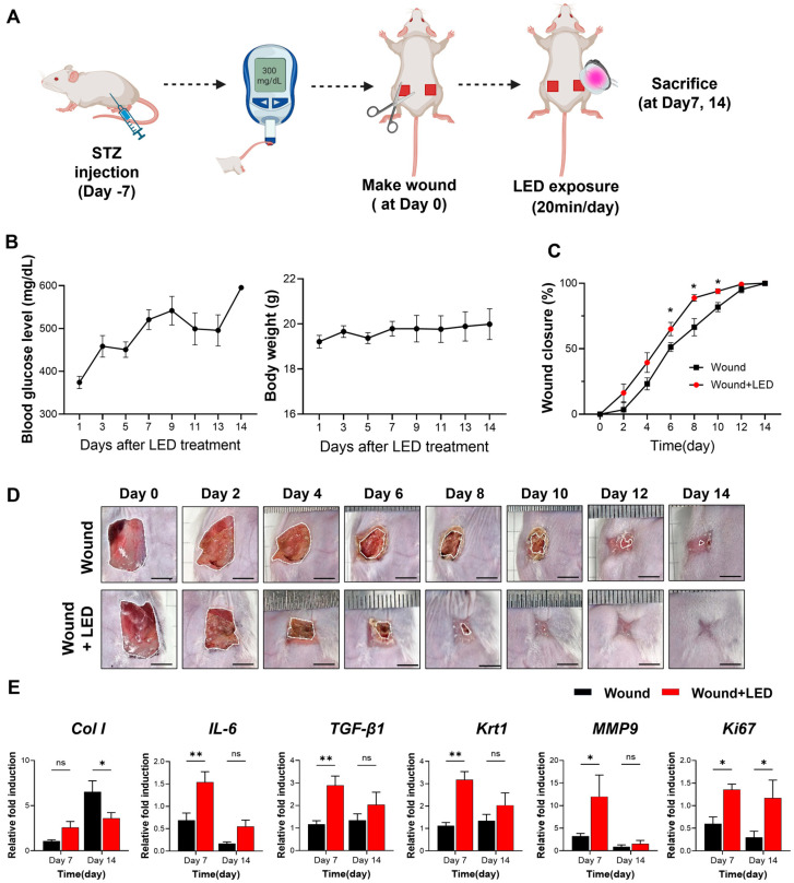
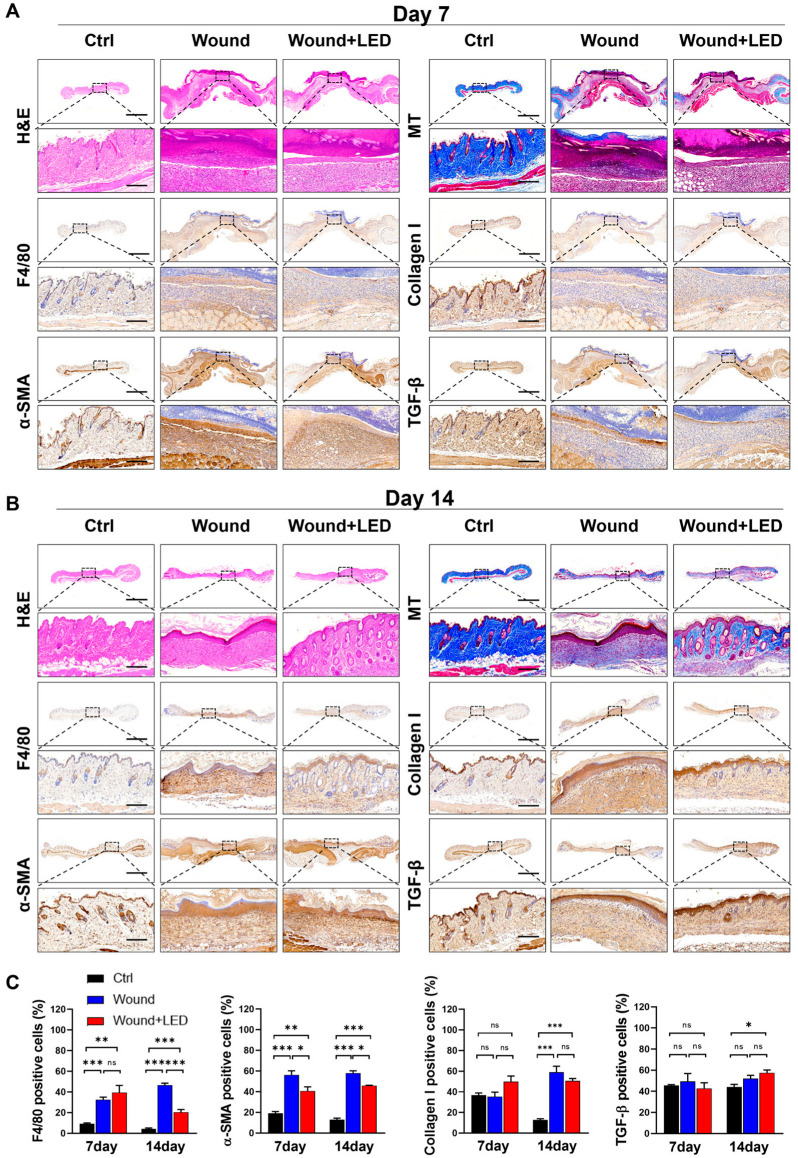
Chronic diabetic wounds affect 15-20% of patients and are characterized by impaired healing due to disrupted hemostasis, inflammation, proliferation, and extracellular matrix (ECM) remodeling. Low-level light therapy (LLLT) has emerged as a promising noninvasive strategy for enhancing tissue regeneration. Here, we developed a multispectral pulsed LED system combining red and near-infrared light to stimulate wound healing. In vitro photostimulation of human keratinocytes and fibroblasts on biomimetic hydrogels enhanced adhesion, spreading, migration, and proliferation via increased focal adhesion kinase (pFAK), paxillin, and F-actin expression. In vivo, daily LED treatment of streptozotocin-induced diabetic wounds accelerated closure and improved ECM remodeling. Histological and molecular analyses revealed elevated levels of MMPs, interleukins, collagen, fibronectin, FGF2, and TGF-β1, supporting regenerative healing without excessive fibrosis. These findings demonstrate that multispectral pulsed photobiomodulation enhances diabetic wound healing through focal adhesion-mediated cell migration and ECM remodeling, offering a cost-effective and clinically translatable approach for chronic wound therapy.

Keywords: cell migration; diabetic wound; extracellular matrix (ECM) remodeling; focal adhesion; low-level light therapy (LLLT).


Conflict of interest statement

The authors declare no competing interests.

Figures

Figure 1
Figure 1
LED stimulation promotes keratinocyte and dermal fibroblast proliferation. (A) Schematic illustration of the LED treatment workflow for cells cultured on polyacrylamide (PAA) gels. (B) Cell viability assay of HaCaT and HDF cells following 24 h multispectral pulsed LED exposure confirms the absence of phototoxicity. (C) Representative Live/Dead fluorescence micrographs of HaCaT and HDF cells cultured on PAA gels, 24 h post-irradiation. Live cells are stained green; dead cells, red. Scale bar = 400 μm. (D) Phase-contrast images showing time-dependent morphological changes in HaCaT and HDF cells over 72 h with and without LED exposure. Scale bar = 110 μm for HaCaT cells and scale bar = 220 μm for HDF cells. (E) Quantitative analysis of cell proliferation via MTT assay at 24 h intervals for 3 days post-LED irradiation. (F,G) Representative images and quantification of EdU incorporation and Ki-67 immunostaining in HaCaT (F) and HDF (G) cells, indicating enhanced DNA synthesis and proliferative activity. EdU-positive nuclei: green; Ki-67-positive nuclei: red; total nuclei: blue. Scale bar = 200 μm. Statistical analysis of HDF cells showing EdU-positive (green) and Ki-67-positive (red) proliferating cells. Nuclei are stained blue. Scale bar = 200 μm. Statistical significance: ns, p > 0.05; * p < 0.05; ** p < 0.01.
Figure 2
Figure 2
LED irradiation enhances the migratory behavior of keratinocytes and dermal fibroblasts. (A,B) Representative trajectories of single HaCaT (A) and HDF (B) cells tracked over a 6 h period on PAA hydrogels, with or without LED stimulation. All tracks are normalized to the origin (0,0). The color gradient indicates migration speed (µm/h). (C,D) Mean squared displacement (MSD) curves showing significantly enhanced displacement of HaCaT (C) and HDF (D) cells following LED treatment. (E,F) Mean absolute distance (MAD) of individual HaCaT (E) and HDF (F) cells over time, further confirming increased motility under LED exposure. (G,H) Quantification of diffusion coefficients and average velocities of HaCaT (G) and HDF (H) cells, derived from MSD and MAD analyses. Statistical significance: ** p < 0.01; *** p < 0.001.
Figure 3
Figure 3
LED irradiation enhances skin cell migration and upregulates pro-healing gene expression. (A,B) Representative phase-contrast images of HaCaT (A) and HDF (B) cells undergoing scratch wound closure on PAA hydrogels over time, with or without LED treatment. White dashed lines mark the initial wound boundary at 0 h. Scale bar = 300 μm. Quantification of wound closure (%) was performed using MATLAB based on the remaining wound area relative to the initial gap width. Statistical significance: ns, p > 0.05; * p < 0.05; ** p < 0.01; *** p ≤ 0.001, determined by Two-way ANOVA (C,D) RT-qPCR analysis of mRNA expression 12 h post-LED irradiation in HaCaT (C) and HDF (D) cells. Genes associated with ECM remodeling (IL-6, MMP-2, MMP-9), ECM synthesis (COL1, COL3, fibronectin), and cell migration (N-cadherin, vimentin) were evaluated. GAPDH served as the internal control. The mean ± Standard Error of the Mean (SEM) (n = 3) values.
Figure 4
Figure 4
LED photostimulation enhances focal adhesion signaling and cytoskeletal organization in HaCaT keratinocytes. (A) Representative immunofluorescence images of HaCaT cells cultured on polyacrylamide (PAA) gels and subjected to LED photostimulation. Cells were stained for phosphorylated FAK (p-FAK, green), paxillin (red), and F-actin (cyan, phalloidin), with nuclei counterstained using Hoechst (blue). Insets highlight focal adhesion complexes and actin architecture. Scale bars = 50 μm, Inlet scale bar = 5 μm. (BD) Quantitative image analysis of focal adhesion and cytoskeletal features—including signal intensity and morphological characteristics of F-actin, p-FAK, and paxillin—was performed using MATLAB (n > 90 cells). Statistical significance: * p < 0.05; ** p < 0.01; *** p < 0.001.
Figure 5
Figure 5
LED photostimulation enhances focal adhesion signaling and cytoskeletal organization in human dermal fibroblasts. (A) Representative immunofluorescence images of HDF cells cultured on polyacrylamide (PAA) gels and subjected to LED photostimulation. Cells were stained for phosphorylated FAK (p-FAK, green), paxillin (red), and F-actin (cyan, phalloidin), with nuclei counterstained using Hoechst (blue). Insets highlight focal adhesion structures and actin fiber alignment. Scale bars = 50 μm, Inlet scale bar = 5 μm. (BD) Quantitative analysis of cytoskeletal and focal adhesion features—including intensity and morphological parameters of F-actin, p-FAK, and paxillin—was performed using MATLAB (n > 90 cells per condition). Statistical significance: * p < 0.05; ** p < 0.01; *** p < 0.001.
Figure 6
Figure 6
LED photobiomodulation accelerates wound healing in an STZ-induced diabetic mouse model. (A) Schematic of the in vivo experimental timeline. Diabetes was induced by streptozotocin (STZ) injection, and full-thickness dorsal wounds were created 7 days later (Day 0). Mice received daily LED photobiomodulation (20 min/day) and were sacrificed on Days 7 or 14 for analysis. (B) Blood glucose levels and body weight were monitored throughout this study to confirm hyperglycemia and general health. (C) Wound closure kinetics over 14 days, showing significantly enhanced healing in the LED-treated group compared to the untreated wound group. (D) Representative macroscopic images of wound healing progression at indicated time points. Scale bars = 5 mm. (E) RT-qPCR analysis of wound tissue on Days 7 and 14, showing expression levels of genes involved in matrix remodeling (Col1, MMP9), inflammation (IL-6), re-epithelialization (Krt1), fibrosis (TGF-β1), and proliferation (Ki67). Expression normalized to GAPDH and Day 0 baseline. Data are presented as mean ± SEM (n = 5–7). Statistical significance is indicated as follows: ns, p > 0.05; * p < 0.05; ** p < 0.01.
Figure 7
Figure 7
LED photobiomodulation improves wound tissue regeneration and remodeling in STZ-induced diabetic mice. (A,B) Histological and immunohistochemical evaluation of skin wound tissues collected on Day 7 (A) and Day 14 (B) post-injury from control, untreated wound, and LED-treated wound groups. Stainings include H&E (epidermal and dermal structure), Masson’s Trichrome (collagen deposition), immunohistochemistry for F4/80 (macrophage infiltration), collagen I (ECM remodeling), α-SMA (myofibroblast activation), and TGF-β1 (fibrotic signaling). Scale bars: 1500 μm (overview panels), 180 μm (insets). (C) Quantification of positive-stained cells from immunohistochemistry on Days 7 and 14. Values represent mean ± SEM. Statistical significance is indicated: ns, p > 0.05; * p < 0.05; ** p < 0.01; *** p < 0.001.

Similar articles

References

    1. Choudhary V., Choudhary M., Bollag W.B. Exploring Skin Wound Healing Models and the Impact of Natural Lipids on the Healing Process. Int. J. Mol. Sci. 2024;25:3790. doi: 10.3390/ijms25073790. - DOI - PMC - PubMed
    1. Diller R.B., Tabor A.J. The Role of the Extracellular Matrix (ECM) in Wound Healing: A Review. Biomimetics. 2022;7:87. doi: 10.3390/biomimetics7030087. - DOI - PMC - PubMed
    1. Wilkinson H.N., Hardman M.J. Wound healing: Cellular mechanisms and pathological outcomes. Open Biol. 2020;10:200223. doi: 10.1098/rsob.200223. - DOI - PMC - PubMed
    1. Werner S., Grose R. Regulation of wound healing by growth factors and cytokines. Physiol. Rev. 2003;83:835–870. doi: 10.1152/physrev.2003.83.3.835. - DOI - PubMed
    1. Vizely K., Wagner K.T., Mandla S., Gustafson D., Fish J.E., Radisic M. Angiopoietin-1 derived peptide hydrogel promotes molecular hallmarks of regeneration and wound healing in dermal fibroblasts. iScience. 2023;26:105984. doi: 10.1016/j.isci.2023.105984. - DOI - PMC - PubMed

MeSH terms

LinkOut - more resources