. 2025 Jun 29;26(13):6281.
doi: 10.3390/ijms26136281.

UBC9-Mediated SUMO Pathway Drives Prohibitin-1 Nuclear Accumulation and PITX1 Repression in Primary Osteoarthritis

Affiliations

UBC9-Mediated SUMO Pathway Drives Prohibitin-1 Nuclear Accumulation and PITX1 Repression in Primary Osteoarthritis

Roxanne Doucet et al. Int J Mol Sci. .

Abstract

Osteoarthritis (OA) is a prevalent and debilitating joint disease in older adults with a complex etiology. We investigated the role of SUMOylation, a post-translational modification, in OA pathogenesis, focusing on the mitochondrial chaperone Prohibitin (PHB1) and the cartilage homeostasis transcription factor PITX1. We hypothesized that oxidative stress-induced SUMOylation promotes PHB1 nuclear accumulation, leading to PITX1 downregulation and contributing to OA development. Analysis of cartilage specimens from 27 OA patients and 4 healthy controls revealed an increased nuclear accumulation of PHB1 in OA chondrocytes, accompanied by elevated levels of SUMO-1 and SUMO-2/3. Mechanistically, nuclear PHB1 interacted indirectly with SUMO-1 through a SUMO-interacting motif (SIM), and the deletion of this SIM prevented PHB1 nuclear trapping in OA cells. Furthermore, the SUMO-conjugating enzyme E2 (UBC9) encoded by the UBE2I gene was upregulated in knee OA cartilage, and its overexpression in vitro enhanced PHB1 nuclear accumulation. Consistently, transgenic mice overexpressing the Ube2i gene exhibited increased UBC9 in their knee cartilage, resulting in Pitx1 downregulation and the emergence of an early OA-like phenotype in articular chondrocytes. Our findings uncover a novel role for UBC9-mediated SUMOylation in primary knee and hip OA. This pathway enhances PHB1 nuclear accumulation, contributing to PITX1 repression and subsequent OA development. These results underscore the importance of SUMOylation in OA pathogenesis and suggest potential molecular targets for early diagnosis and therapeutic intervention.

Keywords: PITX1; Prohibitin-1 (PHB1); SUMOylation; UBC9; osteoarthritis.


Conflict of interest statement

The authors declare no conflict of interest.

Figures
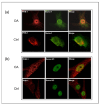
Figure 1
Figure 1
SUMO-1 proteins accumulate in the nuclei of OA articular chondrocytes and co-localize with PHB1. Representative double immunofluorescence staining against PHB1 (red), SUMO-1 (green), and SUMO-2/3 (green) was carried out on articular chondrocytes of one OA patient and one healthy subject. In OA patients, PHB1 shows a distinct accumulation in the nucleus, in contrast to control cells where it is primarily cytoplasmic. Furthermore, the SUMO proteins accumulate in nuclear bodies (NBs) in OA, while control patient cells show little or no accumulation of SUMOs in the nuclear bodies. (a) In OA chondrocytes, PHB1 (red) is co-localized with SUMO-1 (green), appearing as a strong yellow signal in a merged picture (upper panel). In control cells, PHB1 is largely cytoplasmic with minimal nuclear presence and SUMO-1 nuclear bodies are less prominent (lower panel). (b) In OA chondrocytes, a nuclear accumulation of PHB1 (red) is shown, and is co-localized with SUMO-2/3 (green) (upper panel). In control cells, PHB1 remains predominantly cytoplasmic, and SUMO-2/3 nuclear bodies are less defined (lower panel).
Figure 2
Figure 2
SUMO proteins accumulate in PML nuclear bodies in OA articular chondrocytes. Representative double immunofluorescence staining on human articular chondrocytes of OA patients and control subjects was performed with antibodies against PML (green), SUMO-1 (red), and SUMO-2/3 (red). (a) SUMO-1 is co-localized in the PML nuclear bodies of OA chondrocytes. The accumulation of SUMO-1 is in the nucleus, including the PML nuclear bodies. (b) SUMO-2/3 is co-localized in the PML nuclear bodies of OA chondrocytes. In OA chondrocytes, nuclear accumulation of SUMO-2/3 proteins is mainly localized in the PML nuclear bodies.
Figure 3
Figure 3
PML and PHB1 co-localize in OA patients’ nuclei of human articular chondrocytes. Representative double immunofluorescence staining on human articular chondrocytes of OA patients and control subjects was performed with antibodies against PML (green) and PHB-1 (red). The nucleus is stained blue with DAPI. The upper panel (OA) shows that PHB1 accumulates mainly in the nuclei of OA chondrocytes like PML, which colocalizes with the PML nuclear bodies. The lower panel (Ctrl) shows that PHB1 is not detected in the nuclei of control human articular chondrocytes.
Figure 4
Figure 4
In silico analysis of primary amino acid sequences of human PHB1 protein reveals putative SUMOylation sites and SUMO-binding motifs. We used GPS-SUMO (https://sumo.biocuckoo.cn/, accessed on 18 May 2009) for in silico analyses. We selected [all] for SUMOylation and SUMO interaction (SIM) for the threshold. (a) The location of all probabilities of putative SUMOylation associated with each site in PHB1 (red) and for the selected SIM (green). The asterisks (*) indicate amino acids that precede or follow the PHB1 sequence but are not shown. (b) The scheme of SUMOylation sites (blue), SUMO interaction (SIMs) (green), and the selected SUMO-binding site for SIM in PHB1 (red square).
Figure 5
Figure 5
SUMO1 cannot SUMOylate PHB1 in vitro. An in vitro SUMOylation assay in the presence of SUMO-1, E1 and E2 enzymes, ATP, and purified GST-PHB-1 protein indicated that PHB1 could not be SUMOylated in vitro. GST and GST-RanGap1 proteins were used as negative and positive controls, respectively. (a) Four times less GST protein was used for the test than the fusion proteins. The in vitro SUMOylation assay products were analyzed by immunoblots against PHB1 and RanGap1. The asterisk (*) represents the SUMOylated GST-RanGap1. (b) The purified GST and GST fusion proteins were analyzed using SDS-PAGE followed by Coomassie blue staining.
Figure 6
Figure 6
PHB1 can bind SUMO1 proteins via a SIM (SUMO-interacting module), which is crucial for its nuclear localization. (a) Diagram represents wild-type PHB1 protein structure and various PHB1 constructs: wild-type PHB1 (WT PHB1), a mutant where the nuclear signal of export was deleted (PHB1-∆NES) or was replaced by a nuclear localization signal (PHB1-NLS), and a mutant where a putative SUMO-interacting motif (SIM) was deleted (PHB1-∆SIM). All the constructs have a triple Flag-tag at the N-terminal. (b) Co-immunoprecipitation assays with anti-c-Myc antibodies demonstrate that PHB1 interacts with Myc-tagged SUMO1 through the SIM (upper panel). The lower panel indicates the level of Myc-tagged SUMO1 protein in total cell extracts (X-T). (c) The nuclear accumulation of PHB1 is dependent on its SIM. C28/I2, a human chondrocyte cell line, were infected with either flag-tagged wild type (PHB1), (PHB1_NLS), or (PHB1_∆SIM) constructs or empty vector, to produce stable cell lines. The nuclear extract (X-N) and the cytoplasmic extract (X-C) proteins were isolated and analyzed by Western immunoblotting to detect the subcellular presence of flag-tagged PHB1. Anti-GAPDH was used as a cytoplasmic loading control, and anti-Lamin was used as a nuclear loading control. Note the significantly reduced nuclear presence of PHB1-ΔSIM compared to WT PHB1 and PHB1-NLS.
Figure 7
Figure 7
UBC9 expression is increased in the OA cartilage of the knee joint and correlates with the disease severity. (a) Representative immunohistological sections showing UBC9 in the human articular cartilage of control and OA subjects. The left panels show Safranin-O staining (red), which represents the proteoglycan content, which decreases with the severity of OA. The right panels represent IHC staining in superficial and deep zones of normal and OA human articular cartilage, performed with anti-UBC9 antibody (brown signal), where the staining intensity correlates with disease progression. (b) Immunohistochemical quantification of UBC9 in the superficial and deep zones of human cartilage in normal (n = 3) and OA (n = 12). Data are shown as mean ± SEM per condition. * p < 0.05. The scale bar is 100 µm.
Figure 8
Figure 8
The UBC9-mediated SUMO pathway stabilizes PHB1 and promotes its nuclear accumulation in U2OS cells. (a) Co-expression of UBC9 and SUMO isoforms enhances PHB1 protein levels. U2OS cells were transfected with the pLPC-3xFlag-PHB1 alone or co-transfected with different components of the SUMOylation pathway (UBC9; UBC9 + Sumo1; UBC9 + Sumo2; UBC9 + Sumo3). Total cell lysates were analyzed by Western blotting using an anti-Flag antibody to detect Flag-PHB1 protein levels. (b) The nuclear accumulation of PHB1 is dependent on its SIM in the presence of UBC9 and SUMO-1. U2OS cells were transfected with Flag-tagged PHB1, PHB1-NLS, or PHB1-ΔSIM constructs, in the presence or absence of co-transfected Myc-SUMO-1 and UBC9. The nuclear proteins (=X-N), as well as total proteins (X-T), were isolated from cells transfected with pLPC-3xFlag-PHB1, PHB1-NLS, or PHB1-∆SIM in the presence or absence of myc-SUMO1 and UBC9. Anti-GAPDH was used as a cytoplasmic loading control, and anti-Lamin was used as a nuclear loading control, demonstrating successful cell fractionation. (c) U2OS cells were transfected with UBC9 alone or co-transfected with pCMV4-myc-SUMO1, HA-SUMO2, and myc-SUMO3 or with the empty vector. Total (T), cytoplasmic (C), and nuclear (N) protein extracts were isolated. Western blot analysis using an anti-PHB1 antibody reveals changes in endogenous PHB1 subcellular localization. Anti-GAPDH and Anti-Lamin A/C were used as loading controls for cytoplasmic and nuclear fractions, respectively.
Figure 9
Figure 9
X-ray radiographic analysis of knee joints at 35 weeks in wild-type and UBC9-overexpressing transgenic mice. Representative radiographs of knee joints from (a) UBC9-overexpressing transgenic (Tg) and (b) wild-type (WT) mice at 35 weeks of age, obtained using a soft X-ray apparatus (Faxitron), illustrating macroscopic structural changes associated with osteoarthritis. The Tg mouse knee joints exhibit significant alterations compared to WT controls, including increased osteophyte formation (yellow squares) along the fibula, tibial plateau, femur, and tibial intercondylar eminence; increased joint space narrowing (JSN) (red square); obvious signs of subchondral sclerosis (blue arrows); joint bony erosions (green arrows); and subchondral cysts (violet arrows).
Figure 10
Figure 10
Histological and immunohistochemical analysis of knee joints from wild-type and UBC9-overexpressing transgenic mice. (a) Safranin-O and Fast Green Staining: Representative histological images of whole knee joint sections from UBC9 transgenic and wild-type mice at 35 weeks of age stained with Safranin-O (red) to visualize proteoglycan content and Fast Green (counterstain). Note the reduced Safranin-O staining intensity in the articular cartilage of the Tg mouse, indicative of proteoglycan loss and cartilage degradation, compared to the strong and uniform staining in the WT mouse. Scale bar = 100 μm. (b) Immunohistochemistry for UBC9: Representative images showing immunohistochemical staining for UBC9 protein (brown signal) in knee joint sections from Tg and WT mice. The Tg mouse exhibits markedly stronger and more widespread UBC9 staining within chondrocytes compared to the minimal staining observed in the WT control. Scale bar = 100 μm. (c) Immunohistochemistry for Pitx1: Representative images showing immunohistochemical staining for Pitx1 protein (brown signal) in knee joint sections from Tg and WT mice. A substantial downregulation of Pitx1 staining intensity is evident in the chondrocytes of the Tg mouse compared to the more consistent and intense Pitx1 staining in the WT cartilage. Scale bar = 100 μm. All results are representative of experiments conducted with WT (n = 12) and Tg (n = 12) mice.

References

    1. Tang S., Zhang C., Oo W.M., Fu K., Risberg M.A., Bierma-Zeinstra S.M., Neogi T., Atukorala I., Malfait A.-M., Ding C., et al. Osteoarthritis. Nat. Rev. Dis. Primers. 2025;11:10. doi: 10.1038/s41572-025-00594-6. - DOI - PubMed
    1. Primorac D., Molnar V., Rod E., Jeleč Ž., Čukelj F., Matišić V., Vrdoljak T., Hudetz D., Hajsok H., Borić I. Knee Osteoarthritis: A Review of Pathogenesis and State-Of-The-Art Non-Operative Therapeutic Considerations. Genes. 2020;11:854. doi: 10.3390/genes11080854. - DOI - PMC - PubMed
    1. Vidal-Bralo L., Lopez-Golan Y., Mera-Varela A., Rego-Perez I., Horvath S., Zhang Y., del Real Á., Zhai G., Blanco F.J., Riancho J.A., et al. Specific premature epigenetic aging of cartilage in osteoarthritis. Aging. 2016;8:2222–2231. doi: 10.18632/aging.101053. - DOI - PMC - PubMed
    1. Tardio L., Andrés-Bergós J., Zachara N.E., Larrañaga-Vera A., Rodriguez-Villar C., Herrero-Beaumont G., Largo R. O-linked N-acetylglucosamine (O-GlcNAc) protein modification is increased in the cartilage of patients with knee osteoarthritis. Osteoarthr. Cartil. 2014;22:256–263. doi: 10.1016/j.joca.2013.12.001. - DOI - PubMed
    1. Catterall J.B., Hsueh M.F., Stabler T.V., McCudden C.R., Bolognesi M., Zura R., Jordan J.M., Renner J.B., Feng S., Kraus V.B. Protein modification by deamidation indicates variations in joint extracellular matrix turnover. J. Biol. Chem. 2012;287:4640–4651. doi: 10.1074/jbc.M111.249649. - DOI - PMC - PubMed

MeSH terms

LinkOut - more resources