. 2025 Jul 4;26(13):6462.
doi: 10.3390/ijms26136462.

Moloney Murine Leukemia Virus-like Nanoparticles Pseudo-Typed with SARS-CoV-2 RBD for Vaccination Against COVID-19

Affiliations

Moloney Murine Leukemia Virus-like Nanoparticles Pseudo-Typed with SARS-CoV-2 RBD for Vaccination Against COVID-19

Bernhard Kratzer et al. Int J Mol Sci. .

Abstract

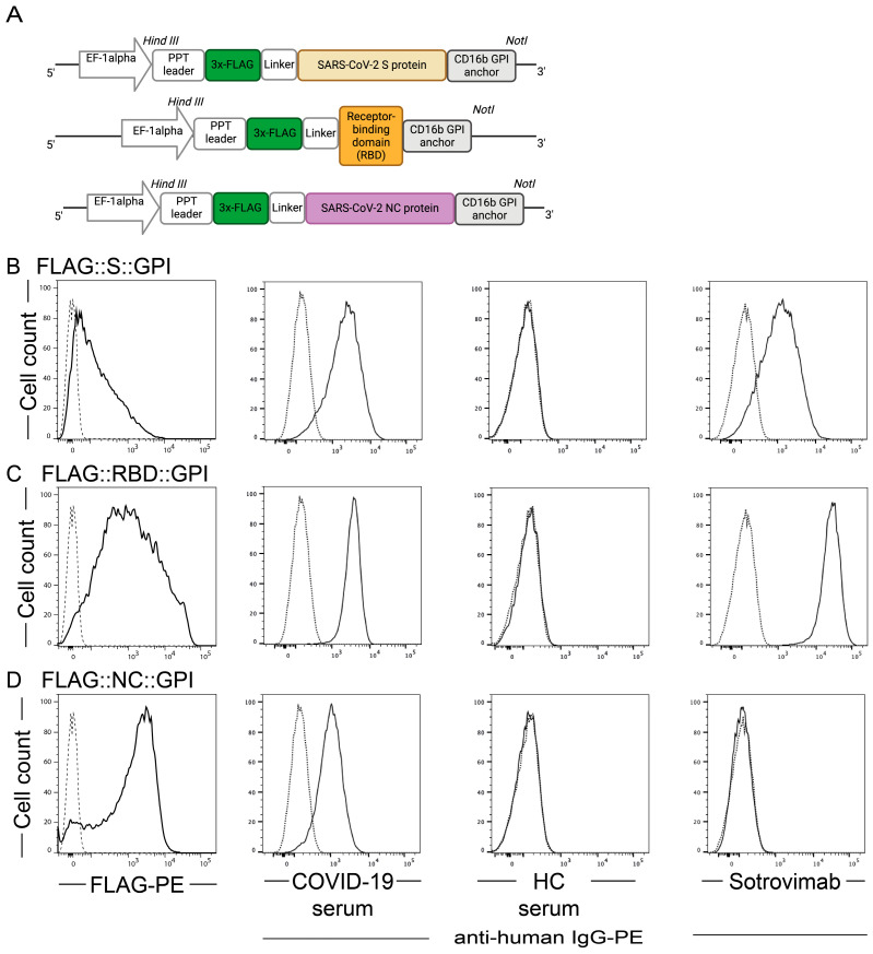
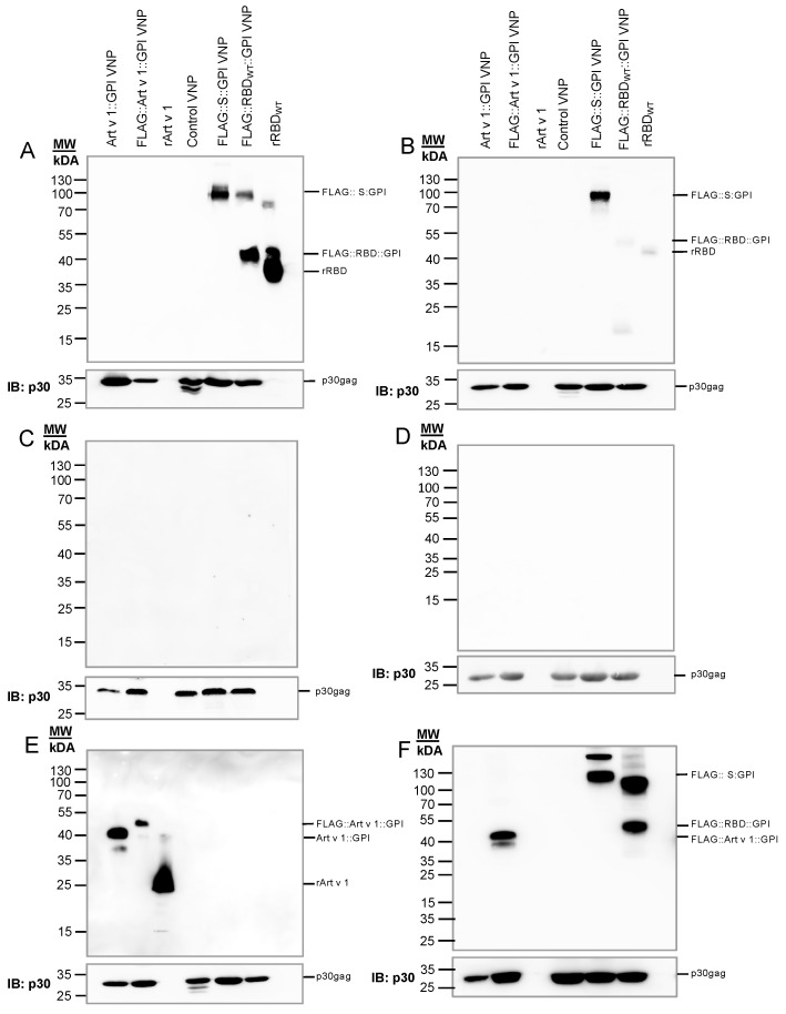
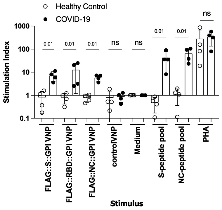
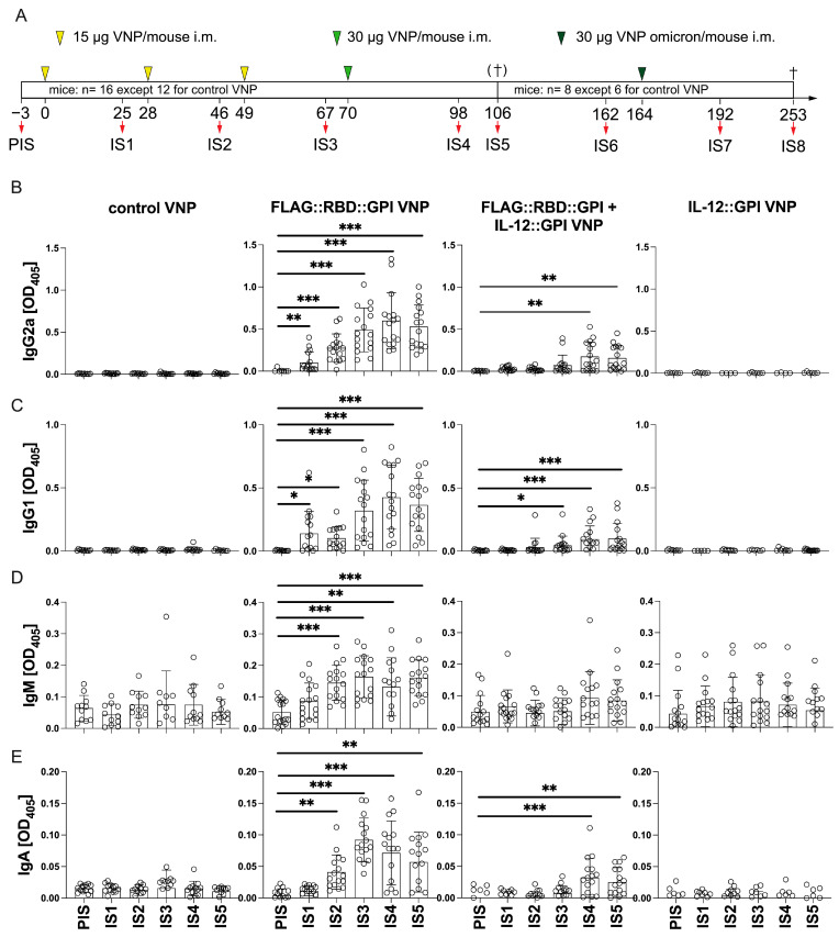
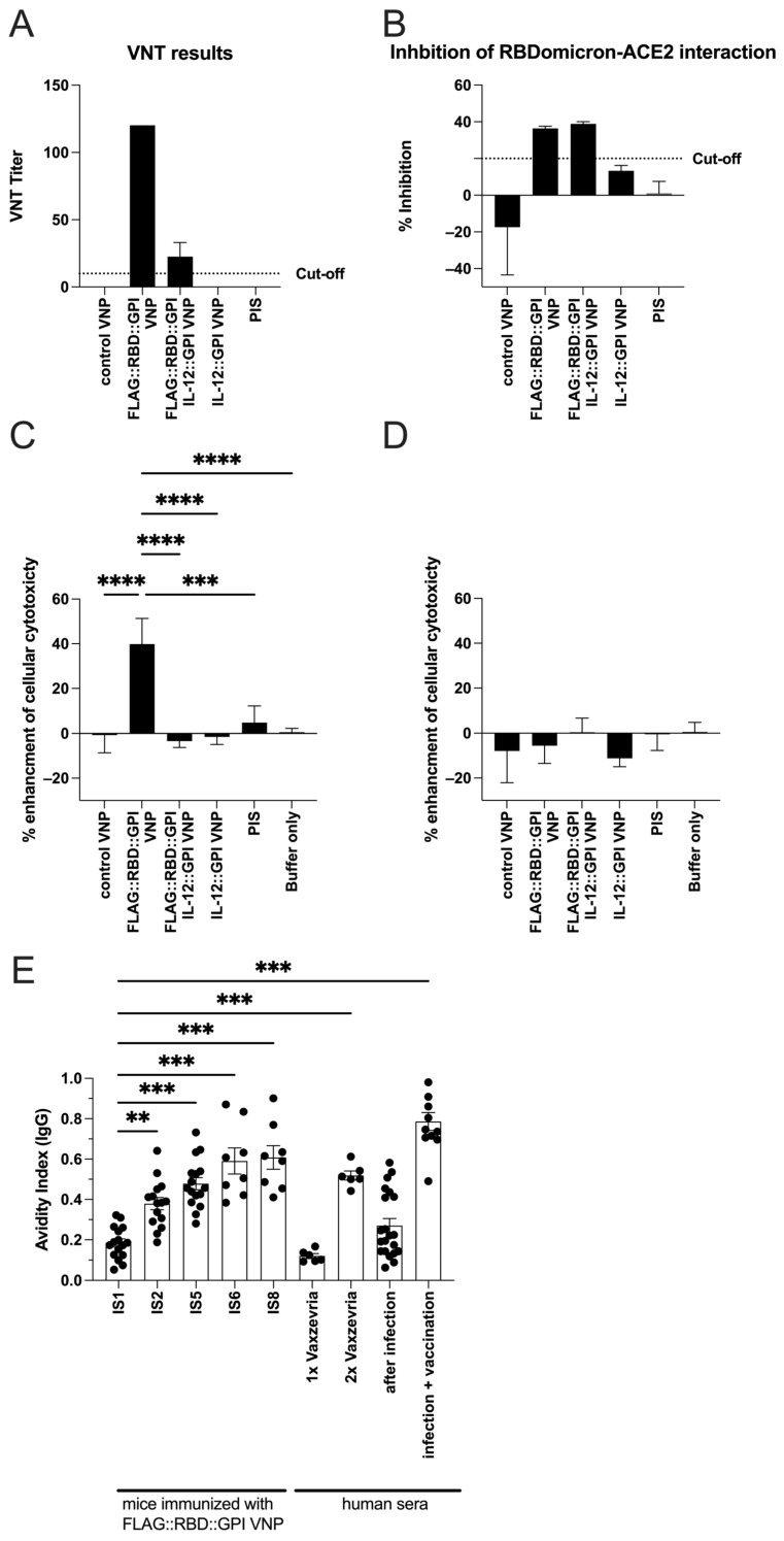
Virus-like nanoparticles (VNPs) based on Moloney murine leukemia virus represent a well-established platform for the expression of heterologous molecules such as cytokines, cytokine receptors, peptide MHC (pMHC) and major allergens, but their application for inducing protective anti-viral immunity has remained understudied as of yet. Here, we variably fused the wildtype SARS-CoV-2 spike, its receptor-binding domain (RBD) and nucleocapsid (NC) to the minimal CD16b-GPI anchor acceptor sequence for expression on the surface of VNP. Moreover, a CD16b-GPI-anchored single-chain version of IL-12 was tested for its adjuvanticity. VNPs expressing RBD::CD16b-GPI alone or in combination with IL-12::CD16b-GPI were used to immunize BALB/c mice intramuscularly and subsequently to investigate virus-specific humoral and cellular immune responses. CD16b-GPI-anchored viral molecules and IL-12-GPI were well-expressed on HEK-293T-producer cells and purified VNPs. After the immunization of mice with VNPs, RBD-specific antibodies were only induced with RBD-expressing VNPs, but not with empty control VNPs or VNPs solely expressing IL-12. Mice immunized with RBD VNPs produced RBD-specific IgM, IgG2a and IgG1 after the first immunization, whereas RBD-specific IgA only appeared after a booster immunization. Protein/peptide microarray and ELISA analyses confirmed exclusive IgG reactivity with folded but not unfolded RBD and showed no specific IgG reactivity with linear RBD peptides. Notably, booster injections gradually increased long-term IgG antibody avidity as measured by ELISA. Interestingly, the final immunization with RBD-Omicron VNPs mainly enhanced preexisting RBD Wuhan Hu-1-specific antibodies. Furthermore, the induced antibodies significantly neutralized SARS-CoV-2 and specifically enhanced cellular cytotoxicity (ADCC) against RBD protein-expressing target cells. In summary, VNPs expressing viral proteins, even in the absence of adjuvants, efficiently induce functional SARS-CoV-2-specific antibodies of all three major classes, making this technology very interesting for future vaccine development and boosting strategies with low reactogenicity.

Keywords: COVID-19; SARS-CoV-2; SARS-CoV-2 immunity; VNP; antibody response.


Conflict of interest statement

With regard to the authors disclosure of potential conflicts of interest we would like to acknowledge that Winfried F. Pickl has received honoraria from Novartis, Astra Zeneca and Roche. Rudolf Valenta has received research grants from HVD Life-Sciences, Vienna, Austria, and from Worg Pharmaceutical, Hangzhou, China. He serves as consultant for HVD. The other authors have no conflict of interest to declare. The authors with a Russian affiliation declare that they have prepared the article in their “personal capacity” and/or that they are employed at an academic/research institution where research or education is the primary function of the entity.

Figures

Figure 1
Figure 1
Schematic depiction of expression cassettes and expression of SARS-CoV-2 proteins by HEK-293T VNP producer cells, as detected by an anti-FLAG-tag antibody, anti-RBD antibody Sotrovimab and human sera. (A) Schematic representation of the SARS-CoV-2 proteins in the expression vector pEAK12. Lower panels show the HEK-293T expression of (B) S-protein, (C) RBD protein and (D) NC-protein, as detected by anti-FLAG mAb (FLAG-PE), by serum of a COVID-19-convalescent patient (COVID-19 serum) in comparison to the staining obtained with serum of a SARS-CoV-2 non-exposed control subject (HC) (all as solid lines) or the monoclonal antibody sotrovimab. The dotted lines represent the fluorescence obtained with control-vector-transduced HEK-293T cells using the indicated primary and secondary staining reagents, indicating the background fluorescence. X-axes show the fluorescence intensities and Y-axes show the cell numbers. Data are representative for three independent experiments performed with the sera of five COVID-19-convalescent patients and five SARS-CoV-2 non-exposed control individuals.
Figure 2
Figure 2
Sera of COVID-19-convalescent subjects recognizing SARS-CoV-2 proteins expressed on immunoblotted VNPs. Immunoblot analyses demonstrating the recognition of SARS-CoV-2 proteins expressed on (VNPs) by a serum pool of COVID-19-convalescent subjects or a non-SARS-CoV-2-exposed subject. Ten µg/lane of purified VNPs expressing the indicated SARS-CoV-2::GPI-anchored fusion proteins, VNPs expressing Art v 1::GPI as control for a non-viral GPI-anchored protein, empty control VNPs or 0.25 µg/lane of recombinant RBD or 0.5 µg/mL of recombinant Art v 1 were separated using 11% SDS-PAGE and transferred onto nitrocellulose membranes. The reactivity of a serum pool from five COVID-19-convalescent individuals with purified VNPs and control proteins which had been separated under (A) non-reducing or (B) reducing conditions (upper panels, respectively) or with a serum from a non-SARS-CoV-2-exposed subject under (C) non-reducing or (D) reducing conditions (upper panels, respectively). (E) Reactivity of an anti-Art v 1 mAb (clone 5) under reducing conditions (upper panel). (F) Reactivity with anti-FLAG mAb under reducing conditions. Below each blot, the reactivity of the rat-anti-p30gag mAb R187 with MoMLV core proteins is shown. The position of molecular mass markers is indicated in kilo Daltons (kDa). Control VNPs were generated by HEK-293T-producer cell transfection with OGP vector and an empty pEAK12 vector. The data shown are representative of three independent experiments.
Figure 3
Figure 3
SARS-CoV-2 fusion protein-expressing VNPs induce specific T-cell proliferations in PBMC of COVID-19-convalescent but not non-SARS-CoV-2-exposed control subjects. Shown are the stimulation indices for PBMC (y-axis) of four COVID-19-convalescent individuals (closed circles) and four non-SARS-CoV-2-exposed control subjects (open circles); which were incubated with the indicated stimuli (x-axis) (VNP preparations (10 µg/mL final concentration), SARS-CoV-2 peptide mixes for S- and NC-protein (150 nM), PHA (10 µg/mL) as positive control or medium alone as negative control). Data are displayed as mean values of triplicates. Each symbol indicates PBMC of one individual. Bars indicate the means of the groups and whiskers indicate the standard deviation in the data. Average counts per minutes in medium alone were 1761 ± 1537. The p-values of the non-normally distributed data were calculated between the two groups for each stimulus independently of the other stimuli using the Mann–Whitney U-test; the corresponding values are shown; ns, not significant.
Figure 4
Figure 4
FLAG::RBD::GPI-decorated VNPs induce RBD-specific antibodies upon i.m. immunization of mice. (A) Shown is the scheme and numbers of mice immunized with the different VNP preparations. Yellow (15 µg) and green (30 µg) arrowheads indicate dose and time points of i.m. administration of VNP preparations. Red arrows indicate the timepoints for blood drawing. PIS, pre-immune serum; IS, immune serum. Figures (BE) show the RBD Wuhan Hu-1-specific IgG2a (B), IgG1 (C), IgM (D) and IgA (E) reactivity (y-axes: OD405 values) of sera (x-axes: PIS to IS5) obtained from mice which were immunized as described on top of the Figure (FLAG::RBD::GPI, FLAG::RBD::GPI plus IL-12::GPI, IL-12::GPI decorated or non-decorated control VNPs) and analyzed by ELISA. Circles show the values of individual mice. p-values were calculated with the Kruskal–Wallis test following Dunn’s multiple comparison test against PIS of each group and are denoted as follows: *, p < 0.05; **, p < 0.01; ***, p < 0.001; only significant differences are shown.
Figure 5
Figure 5
Comparison of the reactivity of mouse immune sera with overlapping peptides of SARS-CoV-2 spike protein (Wuhan Hu-1) and full-length folded or unfolded RBD Wuhan Hu-1; stability of the antibody response and effects of a single RBD–Omicron booster vaccination on the RBD Wuhan Hu-1 and RBD–Omicron-specific antibody levels. (A) IgG reactivity (FIU, fluorescence intensity units, Dyelight 550, y-axis) of pooled sera from 8 VNP-immunized mice, except 6 for control VNP-immunized mice, with micro-arrayed SARS-CoV-2 antigens and S-protein-derived peptides (x-axis). (B) Anti-RBD Wuhan Hu-1 IgG2a and IgG1 antibody reactivity measured by ELISA (y-axes) of mice immunized with either FLAG::RBD::GPI VNPs or FLAG::RBD::GPI+IL-12::GPI VNPs from IS4 to IS8 and PIS (x-axes). (C,D) Anti-RBD-antibody reactivity of sera measured by ELISA (1:500 dilution) against coated RBD Hu-1 (C) or RBD–Omicron (D) at time points IS6 and IS7. Each mouse is represented by a different symbol. IgG2a and IgG1 antibody levels were determined by ELISA and are depicted as OD405 (y-axes). p-values were calculated with the Kruskal–Wallis test following Dunn’s multiple comparison test (B) or Wilcoxon matched rank pairs test (C,D) and denoted as follows: ns, not significant, *, p < 0.05; **, p < 0.01.
Figure 6
Figure 6
VNP-induced antibodies are functionally relevant and their avidity increases upon booster immunizations. (A) Virus neutralization titers (serum dilution, y-axis) of 8 mice per group, except 6 mice for control VNPs, which were tested as two independent pools consisting of 4 or 3 mice, respectively. Mice were immunized four times (IS5) with the indicated VNP preparations (x-axis). (B) Inhibition of RBDomicron to the ACE2 binding of sera from mice immunized five times (IS8) with the indicated VNP preparations (x-axis). The dotted line indicated the cut-off of 20%. Bars indicate means and whiskers indicate standard deviations. (C,D) Enhancement of cellular cytotoxicity of LAK cells against either FLAG::RBD-expressing HEK-293T cells (C) or wildtype HEK-293T cells (D) as targets in the presence of the IS8 from mice which were immunized with the indicated VNP preparations, or in the presence of pre-immune serum (PIS) or buffer only. Bars represent the median ±95% CI enhancement of killing (y-axis). Per immunization group, sera of 8 different mice were analyzed, except 6 for mice immunized with control VNPs in 4 independent ADCC experiments, in which each serum was analyzed in duplicate over 5 different effector–target ratios (40:1, 20:1, 10:1, 5:1, 2.5:1, respectively). The cell concentrations of target cells were kept constant at 1 × 10⁴ cells per well and spontaneous lysis comprised <10% of maximum lysis in each experiment. (E) Avidity index (y-axis) assessed by ELISA of individual immune sera (IS) of RBD–VNP-immunized mice (x-axis) or human Vaxzevria vaccinees at the indicated timepoints for venipuncture and the history of SARS-CoV-2 infection and/or vaccination (x-axis). The results of n = 16 mice for IS1, 2 and 5 are shown; n = 8 mice for IS 6 and 8; n = 6 individuals 22 days after the first and 96–121 days after the second vaccination with Vaxzevria. Bars indicate means and whiskers indicate standard deviations. Every point represents the avidity index for an immunized mouse or a human subject. p-values were calculated with Kruskal–Wallis test following Dunn’s multiple comparison test in (C,D) and one-way ANOVA following Dunnett’s correction for multiple comparison test against IS1 of mice of each group in E and are denoted as follows: **, p < 0.01; ***, p < 0.001 and ****, p < 0.0001; only significant differences are shown.

Similar articles

References

    1. Baden L.R., Rubin E.J. Covid-19–The Search for Effective Therapy. N. Engl. J. Med. 2020;382:1851–1852. doi: 10.1056/NEJMe2005477. - DOI - PMC - PubMed
    1. Lazarus R., Taucher C., Brown C., Corbic Ramljak I., Danon L., Dubischar K., Duncan C.J.A., Eder-Lingelbach S., Faust S.N., Green C., et al. Safety and immunogenicity of the inactivated whole-virus adjuvanted COVID-19 vaccine VLA2001: A randomized, dose escalation, double-blind phase 1/2 clinical trial in healthy adults. J. Infect. 2022;85:306–317. doi: 10.1016/j.jinf.2022.06.009. - DOI - PMC - PubMed
    1. Affonso de Oliveira J.F., Zhao Z., Xiang Y., Shin M.D., Villasenor K.E., Deng X., Shukla S., Chen S., Steinmetz N.F. COVID-19 vaccines based on viral nanoparticles displaying a conserved B-cell epitope show potent immunogenicity and a long-lasting antibody response. Front. Microbiol. 2023;14:1117494. doi: 10.3389/fmicb.2023.1117494. - DOI - PMC - PubMed
    1. Zhang J., Yang W., Huang F., Zhang K. Effectiveness of mRNA and viral-vector vaccines in epidemic period led by different SARS-CoV-2 variants: A systematic review and meta-analysis. J. Med. Virol. 2023;95:e28623. doi: 10.1002/jmv.28623. - DOI - PubMed
    1. Tanriover M.D., Doganay H.L., Akova M., Guner H.R., Azap A., Akhan S., Kose S., Erdinc F.S., Akalin E.H., Tabak O.F., et al. Efficacy and safety of an inactivated whole-virion SARS-CoV-2 vaccine (CoronaVac): Interim results of a double-blind, randomised, placebo-controlled, phase 3 trial in Turkey. Lancet. 2021;398:213–222. doi: 10.1016/S0140-6736(21)01429-X. - DOI - PMC - PubMed

MeSH terms