. 2025 Jul 7;26(13):6533.
doi: 10.3390/ijms26136533.

Identification of a PAK6-Mediated MDM2/p21 Axis That Modulates Survival and Cell Cycle Control of Drug-Resistant Stem/Progenitor Cells in Chronic Myeloid Leukemia

Affiliations

Identification of a PAK6-Mediated MDM2/p21 Axis That Modulates Survival and Cell Cycle Control of Drug-Resistant Stem/Progenitor Cells in Chronic Myeloid Leukemia

Andrew Wu et al. Int J Mol Sci. .

Abstract

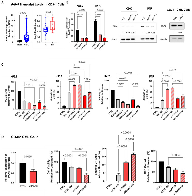
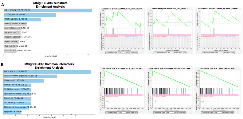
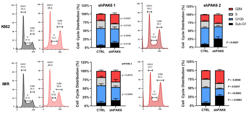
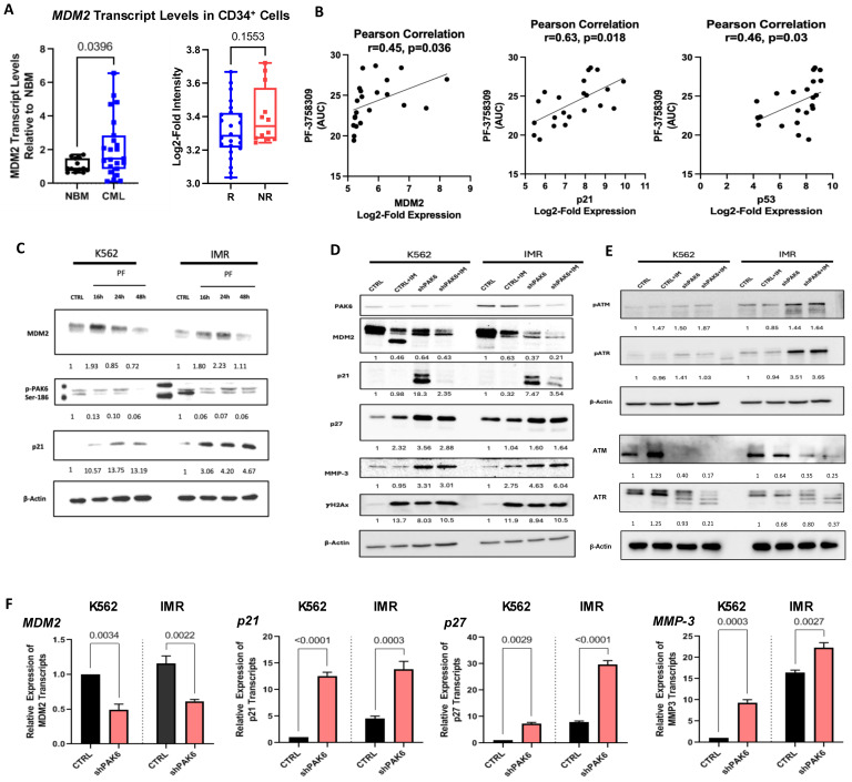
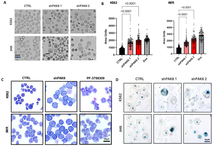
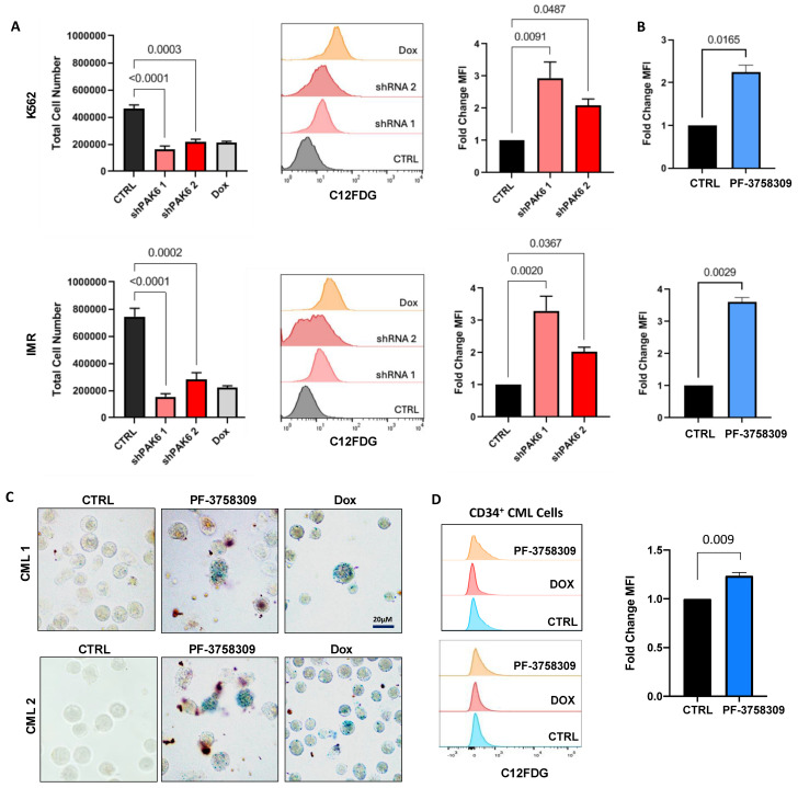
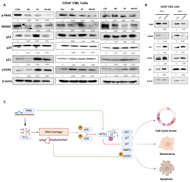
Chronic myeloid leukemia (CML) is a leading example of a malignancy where a molecular targeted therapy revolutionized treatment but has rarely led to cures. Overcoming tyrosine kinase inhibitor (TKI) drug resistance remains a challenge in the treatment of CML. We have recently identified miR-185 as a predictive biomarker where reduced expression in CD34+ treatment-naïve CML cells was associated with TKI resistance. We have also identified PAK6 as a target gene of miR-185 that was upregulated in CD34+ TKI-nonresponder cells. However, its role in regulating TKI resistance remains largely unknown. In this study, we specifically targeted PAK6 in imatinib (IM)-resistant cells and CD34+ stem/progenitor cells from IM-nonresponders using a lentiviral-mediated PAK6 knockdown strategy. Interestingly, the genetic and pharmacological suppression of PAK6 significantly reduced proliferation and increased apoptosis in TKI-resistant cells. Cell survivability was further diminished when IM was combined with PAK6 knockdown. Importantly, PAK6 inhibition in TKI-resistant cells induced cell cycle arrest in the G2-M phase and cellular senescence, accompanied by increased levels of DNA damage-associated senescence markers. Mechanically, we identified a PAK6-mediated MDM2-p21 axis that regulates cell cycle arrest and senescence. Thus, PAK6 plays a critical role in determining alternative cell fates in leukemic cells, and targeting PAK6 may offer a therapeutic strategy to selectively eradicate TKI-resistant cells.

Keywords: CML; DNA damage; Imatinib; PAK6; TKI resistance; TKI therapy; cell cycle; leukemic stem cells; senescence; small molecule inhibitors.


Conflict of interest statement

The authors declare no conflicts of interest.

Figures

Figure 1
Figure 1
Knockdown of PAK6 inhibits cell survival and proliferation. (A) qRT-PCR analysis of PAK6 transcript levels in CD34+ stem/progenitor cells from 23 CML samples compared to 11 normal bone marrow (NBM) samples (left) and analysis of the GSE14671 microarray dataset of CD34+ BM samples from 24 IM-responders and 12 IM-nonresponders (right). (B) Western blot analysis of knockdown of PAK6 with two shRNA constructs in K562 and IMR cells and CD34+ primary CML cells, quantifying protein expression levels relative to β-Actin as indicated. (C) Viability and apoptosis assays were conducted in PAK6 knockdown cells with or without IM. (D) qRT-PCR analysis of PAK6 transcript levels in CD34+ nonresponder cells with PAK6 knockdown (n = 3). Viability, apoptosis, and CFC assays were conducted in these PAK6 knockdown cells with or without IM.
Figure 2
Figure 2
Bioinformatics analysis of PAK6 substrates and common protein interactors. (A,B) Gene set enrichment analyses (GSEAs) of PAK6 substrates and common protein interactors identified from the Harmonizome 3.0 dataset repository revealed significant enrichment of biological processes related to the G2-M checkpoint. * = p value (left). Green line indicate enrichment profile and gray lines indicate raking metric scores (right).
Figure 3
Figure 3
Lentiviral-mediated PAK6 knockdown induces G2/M cell cycle arrest in IM-resistant cells. Flow cytometry analysis of cell cycle distribution in K562 and IM-resistant cells (IMR) following knockdown of PAK6 with two shRNA constructs. PAK6 knockdown reduced the number of cells in the G0/G1 and S phases and induced the accumulation of cells in the G2-M phases compared to the control.
Figure 4
Figure 4
Identification of a PAK6-mediated MDM2-P21 axis in CML cells. (A) qRT-PCR analysis of MDM2 transcript levels in CD34+ cells from 23 CML samples compared to 11 NBM samples (left) and analysis of the GSE14671 microarray dataset from 24 IM-responders and 12 IM-nonresponders (right). (B) Correlation analysis using the Cancer Therapeutics Research Portal (CTRPv2) database between MDM2, p21, and p53 expression and their sensitivities to the PAK inhibitor PF-3758309, as measured by area-under-curve (AUC) values. (C) Western blot analysis of PAK6 phosphorylation, MDM2, and p21 protein expression in K562 and IM-resistant cells (IMR) treated with PF-3758309 (PF) at 16, 24, and 48 h. Quantification of protein expression levels relative to β-Actin as indicated. (DE) Western blot analysis of protein expression and phosphorylation of senescence-associated biomarkers and DNA damage markers, following PAK6 knockdown in K562 and IMR cells, quantifying protein expression levels relative to β-Actin as indicated. (F) qRT-PCR analysis of transcript levels of senescence-associated biomarkers in PAK6 knockdown cells.
Figure 5
Figure 5
Inhibition of PAK6 increases cell size in CML cells. (A,B) Representative phase-contrast images and quantification of cell size in K562 and IM-resistant cells (IMR) following PAK6 knockdown, including senescence-inducing compound doxorubicin (Dox), used as a positive control. (C) GIEMSA staining of K562 and IMR cells following PAK6 knockdown or treatment with PF-3758309 (10 nM). (D) Representative images of senescence-associated β-galactosidase (SABG) staining of K562 and IMR cells following PAK6 knockdown.
Figure 6
Figure 6
PAK6 knockdown induces senescence-associated phenotypes in CML cells. (A) Total cell numbers generated in K562 and IM-resistant cells (IMR) following PAK6 knockdown, including a Dox control, after 48 h of treatment with C12FDG. C12FDG-mediated SABG assays were then performed on these cells and analyzed by FACS histograms, with corresponding quantification of mean fluorescence intensity MFI (bar plots). (B) Fold changes of differences in K562 and IMR cells treated with the PAK inhibitor PF-3758309 (10 nM), with corresponding quantification of MFI by C12FDG-mediated SABG assays. (C,D) Representative images of SABG staining and C12FDG-mediated SABG assays conducted in CD34+ nonresponder primary cells treated with PF-3758309 (10 nM). Intense staining of blue color in cells indicates increased β-galactosidase expression.
Figure 7
Figure 7
PAK6 inhibition elevates senescence-associated and DNA damage response biomarkers in CD34+ CML cells. (A) Western blot analysis of protein expression of senescence-associated biomarkers, including p21, p27, MMP3, and the DNA damage marker γH2Ax, following IM (5µM) or PF-3758309 (10 nM), alone or in combination in CD34+ CML cells (n = 3). (B) Western blot analysis of these proteins in CD34+ CML cells with lentiviral-mediated PAK6 knockdown (n = 2). Quantification of protein expression levels relative to β-Actin as indicated. (C) Model of the role of PAK6 in cell cycle progression, senescence, and DNA damage response in CML. PAK6 promotes leukemia growth by regulating the MDM2-p21 axis, facilitating cell cycle progression, and suppressing senescence. PAK6 knockdown disrupts this axis, leading to G2-M cell cycle arrest, induction of senescence, and activation of the DNA damage response.

Similar articles

References

    1. Sawyers C.L. Chronic Myeloid Leukemia. N. Engl. J. Med. 1999;340:1330–1340. doi: 10.1056/NEJM199904293401706. - DOI - PubMed
    1. Graham S.M., Jørgensen H.G., Allan E., Pearson C., Alcorn M.J., Richmond L., Holyoake T.L. Primitive, Quiescent, Philadelphia-Positive Stem Cells from Patients with Chronic Myeloid Leukemia Are Insensitive to STI571 In Vitro. Blood. 2002;99:319–325. doi: 10.1182/blood.V99.1.319. - DOI - PubMed
    1. Houshmand M., Simonetti G., Circosta P., Gaidano V., Cignetti A., Martinelli G., Saglio G., Gale R.P. Chronic Myeloid Leukemia Stem Cells. Leukemia. 2019;33:1543–1556. doi: 10.1038/s41375-019-0490-0. - DOI - PMC - PubMed
    1. Holyoake T., Jiang X., Eaves C., Eaves A. Isolation of a Highly Quiescent Subpopulation of Primitive Leukemic Cells in Chronic Myeloid Leukemia. Blood. 1999;94:2056–2064. doi: 10.1182/blood.V94.6.2056. - DOI - PubMed
    1. Zhou L.L., Zhao Y., Ringrose A., DeGeer D., Kennah E., Lin A.E.-J., Sheng G., Li X.-J., Turhan A., Jiang X. AHI-1 Interacts with BCR-ABL and Modulates BCR-ABL Transforming Activity and Imatinib Response of CML Stem/Progenitor Cells. J. Exp. Med. 2008;205:2657–2671. doi: 10.1084/jem.20072316. - DOI - PMC - PubMed

MeSH terms

Grants and funding

LinkOut - more resources