. 2025 Jul;38(4):e70038.
doi: 10.1111/pcmr.70038.

Pigmentation and Retinal Pigment Epithelium Thickness: A Study of the Phenotypic and Genotypic Relationships Between Ocular and Extraocular Pigmented Tissues

Affiliations

Pigmentation and Retinal Pigment Epithelium Thickness: A Study of the Phenotypic and Genotypic Relationships Between Ocular and Extraocular Pigmented Tissues

Thomas H Julian et al. Pigment Cell Melanoma Res. 2025 Jul.

Abstract

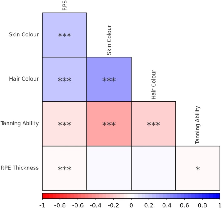
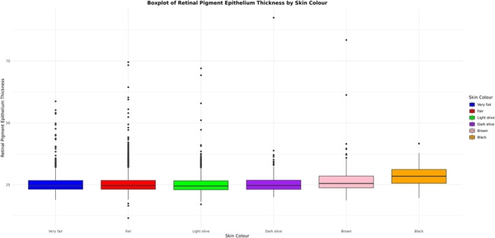
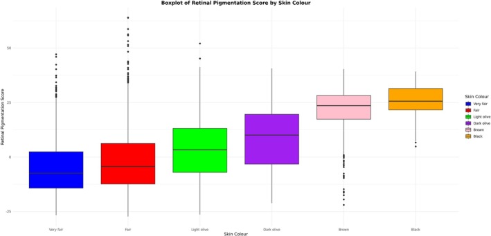
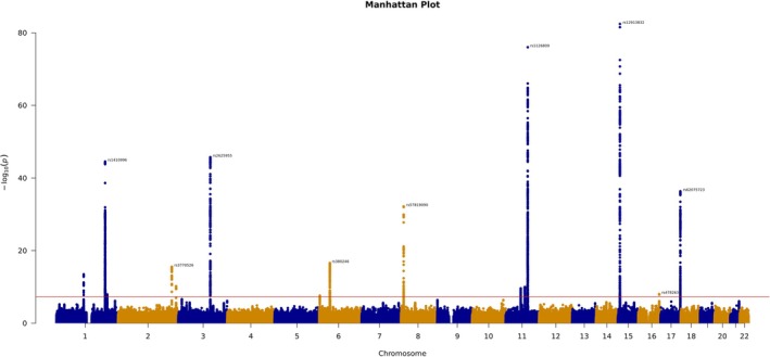
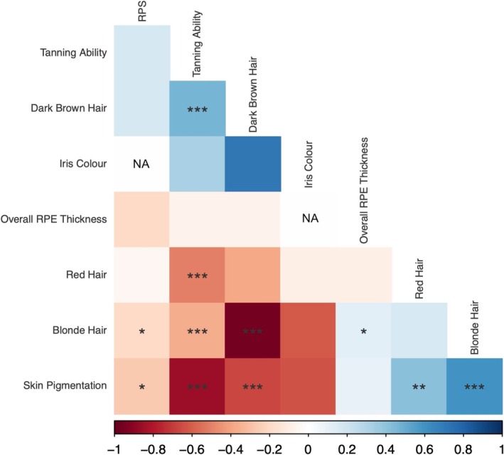
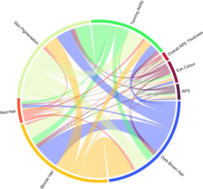
The retinal pigment epithelium (RPE) is a specialised monolayer of pigmented epithelial cells in the outer retina. The extent to which RPE pigmentation is related to that of other tissues remains unclear. We utilised RPE thickness measured using optical coherence tomography (OCT) imaging as an indicator of RPE melanin content. UK Biobank data was used to assess the relationships between RPE thickness and fundus pigmentation, hair colour, skin colour and ability to tan. We performed a genome-wide association study (GWAS) to identify genetic loci associated with RPE thickness. We explored the genetic correlation between RPE thickness and pigmentation-related traits. We found that RPE thickness was not phenotypically or globally genetically correlated with hair colour, skin colour or ability to tan. Whilst RPE thickness was phenotypically correlated with fundus pigmentation, there was not significant global genetic correlation. Despite this, variants in key pigmentation loci including TYR and OCA2-HERC2 were significant in our GWAS of RPE thickness. We identified four genetic regions in which RPE thickness is locally genetically correlated with other pigmentation-related traits, all of which contain protein-coding genes that are central to melanogenesis and melanosome transport. Our study highlights shared and divergent features between RPE thickness and other pigmented traits.

Keywords: GWAS; OCT; RPE; genomics; pigmentation; retina.


Conflict of interest statement

E.B. is a paid consultant and equity holder of Oxford Nanopore, a paid consultant to Dovetail, and a non‐executive director of Genomics England, a limited company wholly owned by the UK Department of Health and Social Care. All other authors declare no conflicts of interest.

Figures

FIGURE 1
FIGURE 1
A phenotypic correlation plot of pigmentation‐related traits available in UK Biobank. Red colour indicates a negative correlation, while blue colour indicates a positive correlation. RPE, retinal pigment epithelium; RPS, retinal pigmentation score. * = p < 0.05, ** = p < 0.01, *** = p < 0.001.
FIGURE 2
FIGURE 2
A box and whisker plot illustrating the distribution of left eye overall average RPE thickness according to self‐reported skin pigmentation.
FIGURE 3
FIGURE 3
A box and whisker plot illustrating the distribution of RPS according to self‐reported skin pigmentation.
FIGURE 4
FIGURE 4
A Manhattan plot, showing the results of a GWAS of left eye overall average RPE thickness, our primary analysis. The red line indicates the threshold for genome‐wide significance and key lead genetic variants above this value have been annotated.
FIGURE 5
FIGURE 5
A correlation plot for the global genetic correlation between pigmentation‐related traits, assessed using LD score regression. Red colour indicates a negative correlation, while blue colour indicates a positive correlation. RPE, retinal pigment epithelium; RPS, retinal pigmentation score. * = p < 0.05, ** = p < 0.01, *** = p < 0.001.
FIGURE 6
FIGURE 6
Chord diagram showing the number of multiple testing corrected statistically significant relationships present between key pigmentation‐related traits. The coloured connections between traits indicate significant relationships; the wider the connecting line, the larger the number of statistically significant local correlations that were identified. Our findings indicate that whilst RPE thickness is genetically correlated with other pigmented tissues in genetic blocks containing pigmentation genes, the overall genetic architecture diverges from that of other tissues. The results suggest that whilst there is not a global correlation between RPE thickness and other pigmented phenotypes, there is a highly significant local correlation in pigment‐specific regions. We wonder if this may in part be due to the differing embryological origins of the pigmented tissues.

Similar articles

Cited by

References

    1. Backman, J. D. , Li A. H., Marcketta A., et al. 2021. “Exome Sequencing and Analysis of 454,787 UK Biobank Participants.” Nature 599, no. 7886: 628–634. - PMC - PubMed
    1. Benito‐Martínez, S. , Zhu Y., Jani R. A., Harper D. C., Marks M. S., and Delevoye C.. 2020. “Research Techniques Made Simple: Cell Biology Methods for the Analysis of Pigmentation.” Journal of Investigative Dermatology 140, no. 2: 257–268.e8. - PMC - PubMed
    1. Boulton, M. , and Dayhaw‐Barker P.. 2001. “The Role of the Retinal Pigment Epithelium: Topographical Variation and Ageing Changes.” Eye 15, no. Pt 3: 384–389. - PubMed
    1. Bouma, B. E. , de Boer J. F., Huang D., et al. 2022. “Optical Coherence Tomography.” Nature Reviews Methods Primers 2: 79. 10.1038/s43586-022-00162-2. - DOI - PMC - PubMed
    1. Bowden, J. , Davey Smith G., Haycock P. C., and Burgess S.. 2016. “Consistent Estimation in Mendelian Randomization With Some Invalid Instruments Using a Weighted Median Estimator.” Genetic Epidemiology 40, no. 4: 304–314. - PMC - PubMed

LinkOut - more resources