. 2025 Jul 12;16(1):518.
doi: 10.1038/s41419-025-07844-1.

O-GlcNAcylation of METTL3 drives hepatocellular carcinoma progression by upregulating MCM10 expression in an m6A-IGF2BP3-dependent manner

Affiliations

O-GlcNAcylation of METTL3 drives hepatocellular carcinoma progression by upregulating MCM10 expression in an m6A-IGF2BP3-dependent manner

Zhen Chen et al. Cell Death Dis. .

Abstract

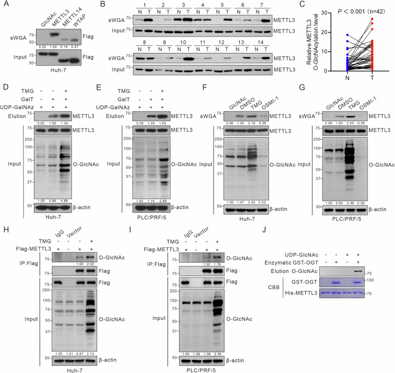
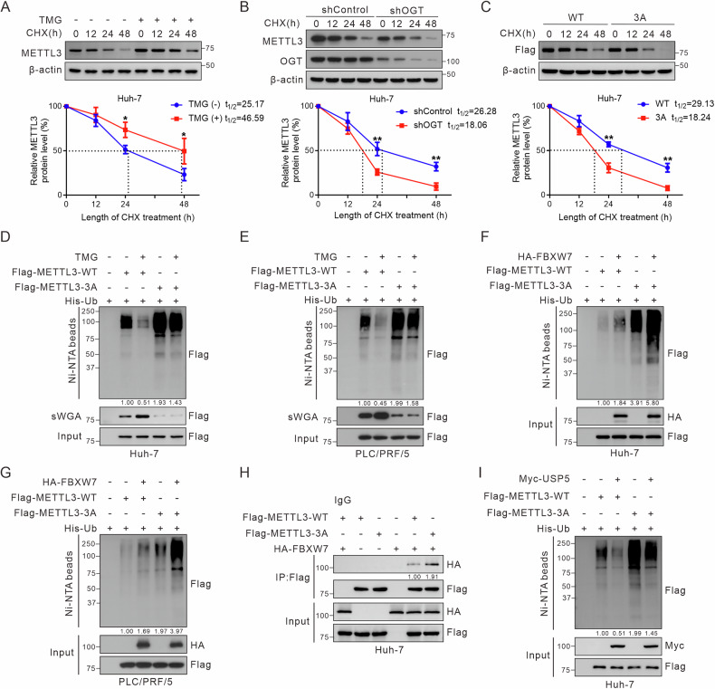
The m6A methyltransferase METTL3 is a key regulator of RNA m6A modification, which plays a critical role in cancer development. Despite the significance of METTL3 in hepatocellular carcinoma (HCC), its post-translational modifications and their functional implications in HCC remain poorly understood. The present study reveals that METTL3 undergoes O-GlcNAcylation, which enhances its stability and promotes HCC progression. Specific O-GlcNAcylation sites (T186/S192/S193) in METTL3 are identified. O-GlcNAc modification reduces METTL3 ubiquitination, thereby increasing protein stability, and enhances its interaction with WTAP, thereby sustaining m6A levels in hepatoma cells. Notably, METTL3 O-GlcNAcylation upregulates the expression of minichromosome maintenance protein 10 (MCM10) by stabilizing its mRNA via an m6A-IGF2BP3-dependent manner. Targeting METTL3 O-GlcNAcylation with designed peptides effectively inhibits HCC growth both in vitro and in vivo. Collectively, our findings provide insights into the regulatory role of O-GlcNAcylation in modulating the m6A epitranscriptome and suggest the potential therapeutic relevance of targeting METTL3 O-GlcNAcylation in HCC.


Conflict of interest statement

Competing interests: The authors declare no competing interests. Ethics approval: All animal experiments were meticulously conducted in compliance with ethics guidelines, and the protocols were approved by the Institutional Animal Care and Use Committee of Chongqing Medical University (approval No: IACUC-CQMU-2023-0185). All clinical tissue sample processing and research activities were performed in strict adherence to the Institutional Review Board protocol (approval No: 2023074).

Figures

Fig. 1
Fig. 1. METTL3 is highly O-GlcNAcylated in HCC.
A Huh-7 hepatoma cells were transfected with Flag-METTL3, Flag-METTL14, or Flag-WTAP for 48 h. Cell lysates were collected for sWGA pull-down assays to identify O-GlcNAcylation. METTL3 O-GlcNAcylation levels in HCC tumors (T) and paired adjacent non-tumor (NT) tissues were detected by sWGA pull-down assays (B), quantified using ImageJ and analyzed with a two-tailed paired Student’s t-test (C), P < 0.001. D, E Analysis of METTL3 O-GlcNAcylation using chemoenzymatic labeling in Huh-7 and PLC/PRF/5 hepatoma cells treated with 25 μM TMG for 12 h. F, G Huh-7 and PLC/PRF/5 cells were treated with 25 μM TMG or 20 μM OSMI-1 for 12 h. Then, cell lysates were pulled down with sWGA-conjugated agarose and immunoblotted with anti-METTL3. H, I Huh-7 and PLC/PRF/5 hepatoma cells were transfected with Flag-METTL3 or a control vector for 48 h. After treatment with 25 μM TMG for 12 h, cell lysates were immunoprecipitated using anti-Flag M2 agarose beads. J For the in vitro O-GlcNAcylation assay, recombinant His-METTL3 proteins and enzymatic GST-OGT domain (aa 313–1031) were incubated in reaction buffer for 4 h. Immunoblot analyses and Coomassie blue staining were performed.
Fig. 2
Fig. 2. OGT mediates O-GlcNAcylation of METTL3 on Thr186/Ser192/Ser193.
A Co-IP assay of the physical interaction between endogenous METTL3 and OGT in Huh-7 cells. B Flag-METTL3 and HA-OGT were transfected into Huh-7 cells. Cell extracts were immunoprecipitated with anti-Flag antibody, followed by immunoblotting with the indicated antibodies. C Pull-down assays were performed to observe the direct interaction between METTL3 and OGT in vitro. Immunoblot analysis and Coomassie blue staining are shown. D Immunofluorescence staining of METTL3 and OGT in Huh-7 cells. Nuclei were counterstained with DAPI. Scale bar: 10 μm. E Schematic representation of the domain structure of METTL3. Full-length METTL3 consists of two domains, a C-terminal region (1–259 aa, ΔC) and the methyltransferase domain (260–580 aa, ΔN). F The interactions between HA-OGT and Flag-METTL3 (WT, ΔC, ΔN) were verified by Co-IP in HEK293 cells. G LC-MS identified Ser193 as the METTL3 O-GlcNAcylation site, which corresponded to O-GlcNAcylated METTL3 peptide AEQDLTTVTTFASSLASGLASSASEPAK. H Cross-species METTL3 sequence alignment. I HEK293 cells were transfected with Flag-tagged METTL3 (WT, T186A, S192A, S193A, or T186A/S192A/S193A). An sWGA pull-down assay was used to identify METTL3 O-GlcNAcylation sites. J, K Huh-7 cells were transfected with Flag-METTL3 WT, 3A mutant, or vector, followed by 25 μM TMG treatment for 12 h. sWGA pull-down (J) and IP (K) assays were used to identify O-GlcNAcylation sites.
Fig. 3
Fig. 3. O-GlcNAcylation of METTL3 at Thr186/Ser192/Ser193 enhances the oncogenic capacity of hepatoma cells in vitro and in vivo.
Huh-7 and PCL/RPF/5 hepatoma cells were infected with METTL3 shRNA lentivirus to decrease endogenous METTL3 levels, followed by infection with adenoviruses expressing Flag-METTL3 (WT or 3A). A Immunoblotting of METTL3 in treated Huh-7 and PCL/PRF/5 hepatoma cells. B, C Growth curves of treated hepatoma cells based on a CCK-8 assay (n = 3 independent experiments). D, E Representative images (100×) and quantitative analysis of cell migration capacity (n = 3, performed in triplicate). Scale bar: 100 μm. F, G Representative images and quantitative results of wound-healing assays of hepatoma cells (100×, n = 3 independent experiments). Scale bar: 200 μm. HJ MHCC-97H cells were processed as described above and injected subcutaneously into nude mice (n = 6 per group). H Representative images of xenograft tumors are shown. Tumor weight (I) and volume (J) were measured and calculated. K, L MHCC-97H cells were treated as described above and injected into nude mice via tail vein injection. Representative images of lung metastases (upper panel) and H&E staining (lower panel) are shown (K) and were quantitatively analyzed (L). Scale bar: 2000 μm. All data are shown as mean ± SD. One-way followed by the Tukey test, *P < 0.05, **P < 0.01, ***P < 0.001.
Fig. 4
Fig. 4. O-GlcNAcylation stabilizes METTL3 by decreasing its ubiquitination.
A Immunoblotting of METTL3. Huh-7 cells were treated with 25 μM TMG for 12 h. Then, 100 μM CHX was added to block protein synthesis for the indicated times. The half-life of METTL3 was quantified in three independent immunoblotting experiments. METTL3 levels at 0 h were arbitrarily set to 100%. B Half-life and quantitative analysis of METTL3. Huh-7 cells were infected with OGT shRNA lentivirus and treated with 100 μM CHX for the indicated times. METTL3 levels were measured by immunoblotting (n = 3). C Half-life of Flag-METTL3 and quantitative analysis in Huh-7 cells. Huh-7 cells were transfected with Flag-METTL3-WT or -3A and treated with 100 μM CHX for the indicated times. Cell lysates were immunoblotted with anti-Flag. Data are representative of three independent experiments. Data in (AC) were analyzed using an unpaired Student’s t-test. *P < 0.05, **P < 0.01. D, E Hepatoma cells were co-transfected with His-Ub and Flag-METTL3 (WT or 3A) and treated or untreated with TMG. Cells were treated with 20 μM MG132 for 8 h, and cell lysates were subjected to IP. Input and IP proteins were immunoblotted with indicated antibodies. FBXW7 increases the poly-ubiquitination level of METTL3 in Huh-7 (F) and PCL/PRF/5 cells (G). H Huh-7 cells were co-transfected Flag-METTL3 (WT or 3A) and HA-FBXW7, cell lysates were subjected to Co-IP to observe their interactions. I USP5 decreases the poly-ubiquitination level of METTL3 in Huh-7 cells.
Fig. 5
Fig. 5. METTL3 O-GlcNAcylation targets MCM10 mRNA to maintain the tumorigenic behavior of hepatoma cells.
A Schematic representation of the METTL3-METTL14-WTAP complex. O-GlcNAcylation of METTL3 enhances its affinity for WTAP, without affecting its association with METTL14. B Flag-METTL3 (WT or 3A) was transfected with or without HA-WTAP into Huh-7 cells. Lysates were immunoprecipitated with anti-Flag antibody, followed by immunoblotting with specific antibodies. C Huh-7-shMETTL3 cells were transfected with Flag-METTL3-WT or -3A. m6A abundance in mRNAs from infected cells was measured using the dot-blot assay with an anti-m6A antibody, with mRNA loading confirmed by methylene blue staining (upper panels). Cell lysates were immunoblotted with the indicated antibodies (lower panels). D Screening for downstream targets of METTL3 by bioinformatic analysis. E Gene Ontology analysis of METTL3-knockdown differentially expressed genes. The mRNA levels of initially screened genes in Huh-7-shMETTL3 cells were measured using RT-qPCR (F), and the m6A modification levels of target mRNAs were assessed using MeRIP-qPCR (G) (n = 3 independent experiments). H Correlation analysis of METTL3 and MCM10 in the TCGA-LIHC cohort (Spearman correlation, P < 0.001). I MeRIP-qPCR assessment of the effect of METTL3 O-GlcNAcylation on m6A modification of MCM10 mRNA. J Immunoblotting to assess the effect of METTL3 O-GlcNAcylation on MCM10 protein levels. KM Huh-7 cells were primarily infected with lentiviruses carrying shControl, shMETTL3, or shMCM10, followed by infection with either Ad-GFP or Ad-MCM10. All treatment groups were subjected to CCK-8 (K), Transwell (L, scale bar: 100 μm), and wound-healing (M, scale bar: 200 μm) assays. Results are derived from three independent experiments and are presented as mean ± SD. Data in (F, G) were analyzed using an unpaired, two-tailed Student’s t-test. Data in (I, KM) were analyzed using one-way ANOVA followed by Tukey tests. *P < 0.05, **P < 0.01, ***P < 0.001.
Fig. 6
Fig. 6. METTL3 O-GlcNAcylation enhances MCM10 mRNA stability in an m6A-IGF2BP3-dependent manner.
A, B RT-qPCR to observe the effect of METTL3 O-GlcNAcylation on relative MCM10 mRNA expression in hepatoma cells. C, D Half-life of MCM10 mRNA in Huh-7 and PLC/PRF/5 cells infected with shMETTL3 lentivirus followed by transfection with Ad-METTL3 (WT or 3A). Transcription was inhibited by actinomycin D (5 μg/mL). E, F Huh-7 and PLC/PRF/5 cells were transfected with shControl or shIGF2BP3 lentivirus. Thirty-six hours after infection, cell lysates were subjected to RT-qPCR to observe MCM10 mRNA expression. G RIP-qPCR assay of MCM10 using IgG or IGF2BP3 antibodies in Huh-7 and PLC/PRF/5 cells. H, I RIP-qPCR assay of MCM10 in Huh-7-shMETTL3-WT (or 3A mutant) and PLC/PRF/5-shMETTL3-WT (or 3A mutant) cells. Half-life of MCM10 mRNA in Huh-7 (J) and PLC/PRF/5 (K) cells infected with shIGF2BP3 lentivirus. L Western blot analysis of MCM10 levels in Huh-7 and PLC/PRF/5 cells infected with IGF2BP3 shRNA. All data represent mean ± SD from three independent experiments. Data in (AF and HK) were analyzed using one-way ANOVA followed by Tukey tests. Data in (G) were analyzed using an unpaired, two-tailed Student’s t-test. **P < 0.01, ***P < 0.001.
Fig. 7
Fig. 7. Targeting METTL3 O-GlcNAcylation suppresses its oncogenic role in HCC.
A Schematic representations of the CPPtat-M1, CPPtat-M2, and CPPtat-M2 mut peptides. Differential residues are highlighted in red. B, C Huh-7 cells were treated with CPPtat, CPPtat-M1, CPPtat-M2, or CPPtat-M2 mut (10 µM) for 24 h, followed by an sWGA pull-down assay to asses METTL3 O-GlcNAcylation. CPPtat-, CPPtat-M2-, CPPtat-M2 mut-treated cells were subjected to CCK-8 (D), colony formation (E), Transwell (F, scale bar: 100 μm), and wound-healing (G, scale bar: 200 μm) assays. Representative images from three independent experiments are shown. H Schematic diagram of the timeline of cell-penetrating peptide (CPPtat, CPPtat-WT, or CPPtat-3A) treatment in the AKT/NRASV12 mouse model. The spontaneous HCC mouse model was established by hydrodynamic tail vein infection. Subsequently, 100 mg/kg of the polypeptides were injected intraperitoneally into mice every 5 days. The schematic was created with BioRender.com (License #RZ28F649PJ, ©2025). I Representative livers of AKT/NRASV12 mice treated with CPPtat, CPPtat-WT, or CPPtat-3A. J The ratio of liver to body weight was calculated (n = 10 mice per group). HCC tissues were harvested for RT-qPCR (K), immunoblotting and sWGA pull-down assays (L). For (J, K), data are presented as mean ± SD, *P < 0.05, ***P < 0.001. M Kaplan–Meier survival curves illustrating the overall survival (OS) of patients with HCC in the TCGA-LIHC cohort according to METTL3 and MCM10 levels, P < 0.05.
Fig. 8
Fig. 8. Schematic model of how METTL3 O-GlcNAcylation promotes HCC progression.
O-GlcNAcylation of METTL3 enhances its stability and interaction with WTAP, upregulating MCM10 mRNA levels in an m6A-IGF2BP3-dependent manner. Created with BioRender.com (License #VY28F5EEOY, ©2025).

References

    1. Fang F, Wang X, Li Z, Ni K, Xiong C. Epigenetic regulation of mRNA N6-methyladenosine modifications in mammalian gametogenesis. Mol Hum Reprod. 2021;27:gaab025. - PubMed
    1. Linder B, Grozhik AV, Olarerin-George AO, Meydan C, Mason CE, Jaffrey SR. Single-nucleotide-resolution mapping of m6A and m6Am throughout the transcriptome. Nat Methods. 2015;12:767–72. - PMC - PubMed
    1. Meyer KD, Saletore Y, Zumbo P, Elemento O, Mason CE, Jaffrey SR. Comprehensive analysis of mRNA methylation reveals enrichment in 3′ UTRs and near stop codons. Cell. 2012;149:1635–46. - PMC - PubMed
    1. Liu J, Yue Y, Han D, Wang X, Fu Y, Zhang L, et al. A METTL3-METTL14 complex mediates mammalian nuclear RNA N6-adenosine methylation. Nat Chem Biol. 2014;10:93–95. - PMC - PubMed
    1. Ping XL, Sun BF, Wang L, Xiao W, Yang X, Wang WJ, et al. Mammalian WTAP is a regulatory subunit of the RNA N6-methyladenosine methyltransferase. Cell Res. 2014;24:177–89. - PMC - PubMed

MeSH terms