. 2025 Jul 12;15(1):25246.
doi: 10.1038/s41598-025-11046-2.

CX3CL1 promotes M1 macrophage polarization and osteoclast differentiation via NSUN5-mediated m5C modification

Affiliations

CX3CL1 promotes M1 macrophage polarization and osteoclast differentiation via NSUN5-mediated m5C modification

Zhibin Deng et al. Sci Rep. .

Abstract

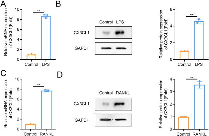
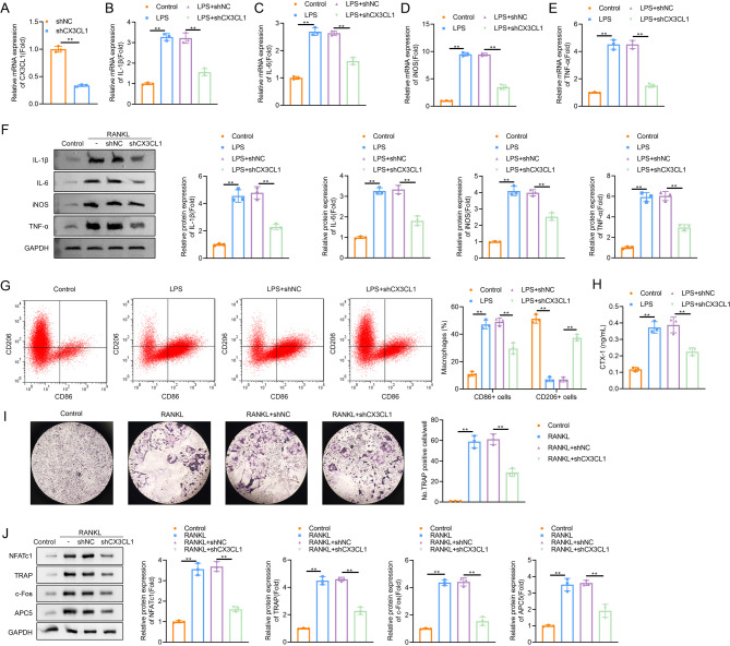
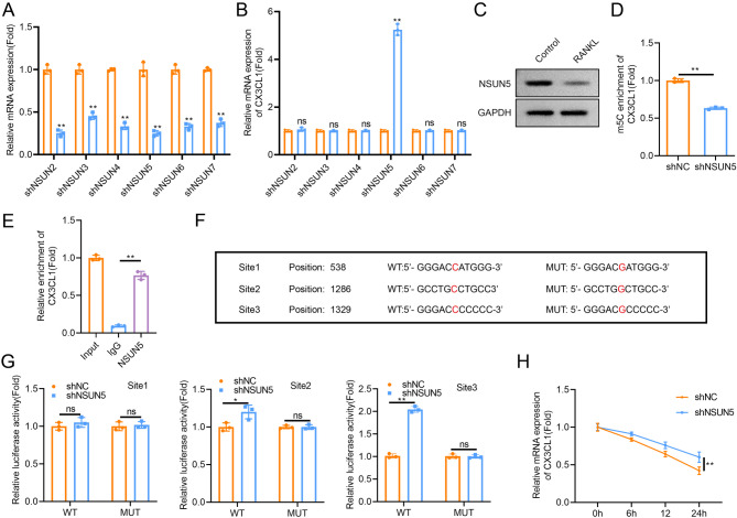
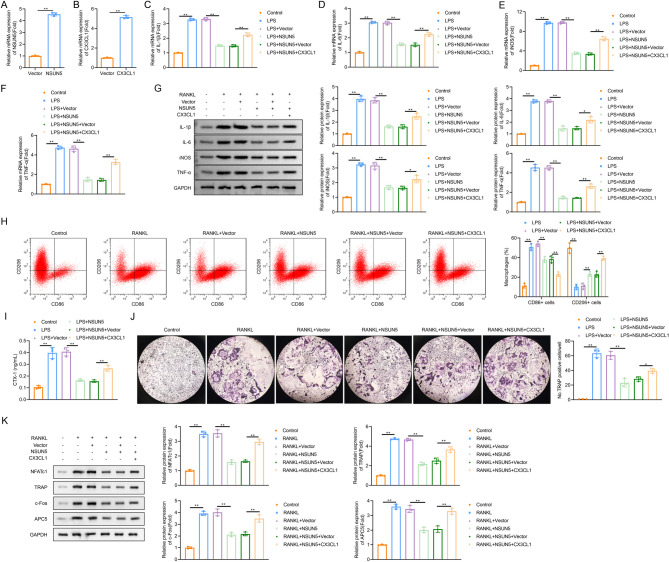
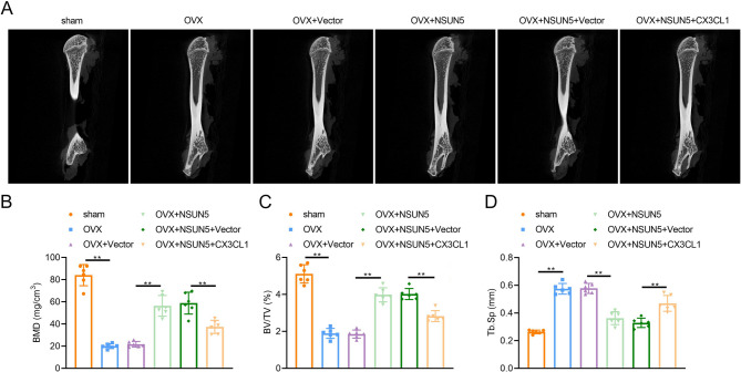
Bone homeostasis refers to a dynamic equilibrium maintained between osteogenesis and osteoclastic bone resorption within the skeletal system. CX3CL1 (Fractalkine) is a chemokine that plays a significant regulatory role in bone homeostasis. This study aimed to investigate the mechanisms by which CX3CL1 regulates bone homeostasis. The expression of CX3CL1 in LPS-stimulated and RANKL-stimulated macrophages was examined using qPCR and Western blotting. Functional studies employed shRNA-mediated knockdown and overexpression of CX3CL1/NSUN5, followed by analysis of pro-inflammatory factor levels(IL-1β, IL-6, iNOS, and TNF-α), M1/M2 markers (CD86/CD206), osteoclast activity (TRAP staining, CTX-1 level), and key osteoclastogenic factors (NFATc1, c-Fos). Potential mechanisms were validated using Methylated RNA Immunoprecipitation (MeRIP), RNA Immunoprecipitation (RIP), and Dual-Luciferase Reporter Assay experiments. An ovariectomy (OVX)-induced osteoporosis mouse model was used for in vivo validation. Results showed that CX3CL1 was significantly upregulated in LPS- and RANKL-stimulated RAW 264.7 cells. Knockdown of CX3CL1 inhibited macrophage M1 polarization and osteoclast differentiation. NSUN5 interacted with CX3CL1 and suppressed its stability by promoting the m5C modification of CX3CL1 mRNA. Additionally, Overexpression of CX3CL1 reversed the inhibitory effect of NSUN5 overexpression on macrophage M1 polarization and osteoclast differentiation. In OVX mice, NSUN5 overexpression preserved bone mass (increased BV/TV, reduced Tb.Sp), while CX3CL1 co-expression abolished this protection. In conclusion, CX3CL1 accelerates M1 macrophage polarization and promotes osteoclast differentiation, mechanistically regulated by m5C modification mediated by NSUN5. This study provides novel therapeutic strategies and targets for maintaining bone homeostasis and preventing and treating bone-related diseases.

Keywords: Bone homeostasis; CX3CL1; M1 polarization; NSUN5; Osteoclast differentiation.


Conflict of interest statement

Declarations. Ethics approval and consent to participate: This study was approved by the Ethics Committee of Songgang People’s Hospital. All animal experiments were complied with the ARRIVE guidelines. All methods were carried out in accordance with relevant guidelines and regulations. Consent for publication: Not applicable. Competing interests: The authors declare no competing interests.

Figures

Fig. 1
Fig. 1
CX3CL1 was highly expressed in vitro. (A-B) CX3CL1 expression was assessed in LPS-stimulated RAW 264.7 cells after 24-h treatment using qPCR for mRNA levels and Western blot analysis for protein expression. (C-D) CX3CL1 expression was assessed in RANKL-stimulated RAW 264.7 cells after 5-days treatment using qPCR for mRNA levels and Western blot analysis for protein expression. Data were analyzed by Student’s t-test. All data are presented as the mean ± SD. **p < 0.01. (n = 3).
Fig. 2
Fig. 2
Knockdown of CX3CL1 inhibited macrophage M1 polarization and osteoclast differentiation. (A) The transfection efficiency was measured by qPCR. (B-F) The mRNA levels and protein expression of IL-1β, IL-6, iNOS, and TNF-α in LPS-stimulated RAW 264.7 cells were detected using qPCR and western blot after CX3CL1 knockdown, respectively. (G) Flow cytometry analysis of CD86+ and CD206+ macrophage proportion in each group. (H) The level of CTX-1 was detected by ELISA assay. (I) The ability of mature osteoclast formation was analyzed by TRAP staining. (J) The expressions of NFATc1, TRAP, c-Fos, and APC5 in RANKL-stimulated RAW 264.7 cells were analyzed by western blot after CX3CL1 knockdown. Data were analyzed by Student’s t-test (A) or one-way ANOVA followed by Tukey’s post hoc test (B-J). All data are presented as the mean ± SD. **p < 0.01. (n = 3).
Fig. 3
Fig. 3
NSUN5 negatively regulated CX3CL1 expression in m5C modification manner. (A) The transfection efficiency was measured by qPCR. (B) The levels of CX3CL1 mRNA were measured using qPCR. (C) The protein expression of NSUN5 in RANKL-stimulated RAW 264.7 cells were measured using Western blot. (D) The m5C modification level of CX3CL1 was detected by MeRIP. (E) The interaction between NSUN5 and CX3CL1 was measured by RIP. (F) The prediction of m5C modification sites in CX3CL1. (G) The binding relationship between NSUN5 and CX3CL1 was analyzed by Luciferase assay. (H) The stability of CX3CL1 mRNA was measured by qPCR after RAW 264.7 cells were treated with actinomycin D at 0, 6, 12, and 24 h. Data were analyzed by Student’s t-test (A, B, D, G), one-way ANOVA (E) or two-way ANOVA (H). All data are presented as the mean ± SD. *p < 0.05 and **p < 0.01. (n = 3).
Fig. 4
Fig. 4
Overexpression of CX3CL1 reversed the inhibitory effect of NSUN5 overexpression on the macrophage M1 polarization and osteoclast differentiation in RAW 264.7 cells. (A-B) The transfection efficiency was analyzed using qPCR. (C-G) The mRNA levels of IL-1β, IL-6, iNOS, and TNF-α in LPS-stimulated RAW 264.7 cells were detected using qPCR. (H) Flow cytometry analysis of CD86+ and CD206+ macrophage proportion in each group. (I) The level of CTX-1 was detected by ELISA assay. (J) The ability of mature osteoclast formation was analyzed by TRAP staining. (K) The expressions of NFATc1, TRAP, c-Fos, and APC5 in RANKL-stimulated RAW 264.7 cells were analyzed by western blot. Data were analyzed by Student’s t-test (A-B) or one-way ANOVA followed by Tukey’s post hoc test (C-K). All data are presented as the mean ± SD. **p < 0.01. (n = 3).
Fig. 5
Fig. 5
CX3CL1 abrogated the bone-protective effects of NSUN5 in OVX-induced osteoporotic mice. (A-B) The effects of CX3CL1 and NSUN5 on (A) Micro-CT, (B) bone mineral density (BMD), (C) bone volume fraction (BV/TV), and (D) trabecular separation (Tb.Sp) of femurs. Data were analyzed by one-way ANOVA followed by Tukey’s post hoc test (B-D). All data are presented as the mean ± SD. **p < 0.01. (n = 6).

Similar articles

References

    1. Choi, J. Y. Healthy bone tissue homeostasis. Exp. Mol. Med.52, 1165. 10.1038/s12276-020-0472-3 (2020). - PMC - PubMed
    1. Chandra, A. & Rajawat, J. Skeletal aging and osteoporosis: mechanisms and therapeutics. Int. J. Mol. Sci.2210.3390/ijms22073553 (2021). - PMC - PubMed
    1. Kong, L., Wang, Y., Smith, W. & Hao, D. Macrophages in bone homeostasis. Curr. Stem Cell. Res. Ther.14, 474–481. 10.2174/1574888X14666190214163815 (2019). - PubMed
    1. Yan, Q., Liu, H., Zhu, R. & Zhang, Z. Contribution of macrophage polarization in bone metabolism: A literature review. Cytokine184, 156768. 10.1016/j.cyto.2024.156768 (2024). - PubMed
    1. Ju, C. et al. Mesenchymal stem cell-associated LncRNA in osteogenic differentiation. Biomed. Pharmacother. 115, 108912. 10.1016/j.biopha.2019.108912 (2019). - PubMed

LinkOut - more resources