. 2025 Dec;14(1):2529632.
doi: 10.1080/2162402X.2025.2529632. Epub 2025 Jul 12.

TIGIT blockade in the context of BCMA-CART cell therapy does not augment efficacy in a multiple myeloma mouse model

Affiliations

TIGIT blockade in the context of BCMA-CART cell therapy does not augment efficacy in a multiple myeloma mouse model

Aina Oliver-Caldes et al. Oncoimmunology. 2025 Dec.

Abstract

BCMA-directed CAR-T therapies have shown promising results in multiple myeloma (MM). However, patients continue to relapse. T cell exhaustion with increased TIGIT expression is a resistance mechanism which was confirmed in CAR-T cells from ARI0002h trial, an academic CAR-T developed in our institution. We aimed to analyze the impact of blocking TIGIT on the efficacy of ARI0002h. We used three different strategies to block TIGIT: (1) Addition of an external blocking anti-TIGIT-antibody (Ab), (2) Modify ARI0002h into a 4th generation CAR-T, named ARITIGIT, capable of secreting a soluble TIGIT-blocking scFv and (3) TIGIT knock-out in ARI0002h using CRISPR/Cas9. Each strategy was evaluated in vitro and in vivo. Adding a TIGIT-blocking Ab to ARI0002h improved in vitro cytotoxicity, but failed to enhance mice survival. The new 4th generation CAR-T, ARITIGIT, was also unable to achieve better survival outcomes despite favoring the in vivo model by using a myeloma cell line with high expression of the TIGIT ligand PVR. Interestingly, when mice were challenged with a second infusion of tumor cells, mimicking a relapse model, a trend for improved survival with ARITIGIT was observed (p = 0.11). Finally, TIGIT-knock-out on ARI0002h (KO-ARI0002h) using CRISPR/Cas9 showed similar in vitro activity to ARI0002h. In an in vivo stress model, TIGIT KO-ARI0002h prolonged survival (p = 0.02). However, this improvement was not significant compared to ARI0002h (p = 0.07). This study failed to demonstrate a significant benefit of TIGIT-blockade on ARI0002h cells despite using three different approaches, suggesting that targeting a single immune checkpoint may be insufficient.

Keywords: 4th generation CAR-T; BCMA-CAR-T; CAR-T cell therapy; CRISPR/Cas9; TIGIT blockade; multiple myeloma.


Conflict of interest statement

AOC declares no conflict of interest. JMP declares no conflict of interest. AMB declares no conflict of interest. LPA declares being one of the inventors of ARI0002h. MB declares no conflict of interest. HC declares no conflict of interest. MC declares no conflict of interest. JC declares no conflict of interest. SVS declares no conflict of interest. MBo declares no conflict of interest. OC declares no conflict of interest. DFM declares no conflict of interest. LGRL declares receiving honoraria and travel grants from Janssen, Amgen, BMS, GSK, Menarini, and Sanofi. EL declares no conflict of interest. LR declares honoraria from Janssen, BMS/Celgene, Amgen, Takeda, Sanofi, and GSK; participation on data safety monitoring or advisory board of Janssen, BMS-Celgene, Amgen, Takeda, Sanofi, and GSK. MJ declares receiving research or travel support by Fundació Bancaria la Caixa, ISCIII, and CellNex Teleom; honoraria from educational activities, speaker, and advisory roles with Miltenyi and indirectly with sponsors of congresses; and participation on data safety monitoring or advisory boards with MAB Gyala. BMA declares being one of the inventors of ARI0002h. CFL declares receiving grants through his institution from BMS, GSK, Janssen, and Amgen.

Figures

Figure 1.
Figure 1.
(a) TIGIT-blockade approaches on the BCMA-directed CAR-T ARI0002h tested in this study. (b) ARITIGIT 4th generation CART sequence scheme.
Figure 2.
Figure 2.
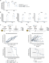
(a) Cytotoxicity results after co-culture of the MM cell lines ARP-1 and U266 with either ARI0002h or untransduced (UT) T cells obtained from healthy donors, adding or not an anti-TIGIT-Ab (the Ab carrier was added to the control groups). Each dot represents a T cell donor; 3 repeats were performed for each condition in each donor. The number of T-cell donors was: (1) ARP1 + UT vs ARP1 + UT + TIGITAb: T cells from n = 8 healthy donors; (2) ARP1 + ARI0002h vs ARP1 + ARI0002h + TIGITAb: T cells from n = 6 healthy donors; (3) U266 + UT vs U266 + UT + TIGITAb: T cells from n = 7 healthy donors; (4) U266 + ARI0002h vs U266 + ARI0002h + TIGITAb T cells from n = 6 healthy donors. Paired students’ t-test and the Wilcoxon ranked sign test were applied according to normality adherence. (b) Pro-inflammatory cytokine production of IFN-γ, IL-2 and TNF-α after 24 hours of co-culture. Each dot represents a T-cell donor; 3 repeats were performed for each condition in each donor. The number of T cell donors was: (1) ARP1 + UT vs ARP1 + UT + TIGITAb: T cells from n = 7 healthy donors; (2) ARP1 + ARI0002h vs ARP1 + ARI0002h + TIGITAb: T cells from n = 6 healthy donors; (3) U266 + UT vs U266 + UT + TIGITAb: T cells from n = 6 healthy donors; (4) U266 + ARI0002h vs U266 + ARI0002h + TIGITAb T cells from n = 5 healthy donors. Paired students’ t-test and the Wilcoxon ranked sign test were applied according to normality adherence. (c) In vivo results of this strategy: mice were administered 1.5x10⁶ MM ARP-1 cells on day 0, 24 h after radiation with 2 Gy. When disease luminescence was detectable, mice were injected with 0.5x10⁶ of a healthy donor-derived ARI0002h CAR+ cells or UT T cells. After that, the anti-TIGIT-Ab was administered intraperitoneally twice a week for 4 weeks in the respective groups. Mice were followed weekly using bioluminescence and sacrifice was performed when prespecified criteria were met. The number of mice in each group was: (1) UT only n = 4; (2) UT + TIGITAb n = 5; (3) ARI0002h n = 5; (4) ARI0002h + TIGITAb n = 5. To analyze survival the log-rank test was applied and survival curves were obtained using the Kaplan-Meier method. (d) In vivo results of the favored model: mice were administered 1.5x10⁶ MM U266 cells on day 0, 24 h after radiation with 2 Gy. When disease luminescence was detectable, mice were injected with 1.5x10⁶ of patient-derived ARI0002h CAR+ cells or UT T cells. After that, the anti-TIGIT-Ab was administered intraperitoneally three times a week, indefinitely, in the respective groups. Mice were followed weekly using bioluminescence and sacrifice was performed when prespecified criteria were met. The number of mice in each group was: (1) UT only n = 2; (2) ARI0002h n = 6; (3) ARI0002h + TIGITAb n = 7. To analyze survival the log-rank test was applied and survival curves were obtained using the Kaplan-Meier method.
Figure 3.
Figure 3.
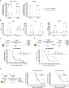
(a) Cytotoxicity results after co-culture of the MM cell lines ARP-1 and U266 with either ARI0002h or ARITIGIT manufactured from healthy donor or patient T cells. Each dot represents a T cell donor; 3 repeats were performed for each condition in each donor. The number of T-cell donors was: (1) ARP1 + ARI0002h vs ARP1 + ARITIGIT + TIGITAb T cells from n = 7 healthy donors; (2) U266 + ARI0002h vs U266 + ARITIGIT + TIGITAb T cells from n = 9 healthy donors; (3) ARP1 + ARI0002h vs ARP1 + ARITIGIT + TIGITAb T cells from n = 4 patients; (4) U266 + ARI0002h vs U266 + ARITIGIT + TIGITAb T cells from n = 4 patients. Paired students’ t-test and the Wilcoxon ranked sign test were applied according to normality adherence. (b) Pro-inflammatory cytokine production of IFN-γ, IL-2 and TNF-α after 24 hours of co-culture. Each dot represents a T cell donor; 3 repeats were performed for each condition in each donor. The number of T-cell donors was: (1) ARP1 + ARI0002h vs ARP1 + ARITIGIT + TIGITAb T cells from n = 7 healthy donors; (2) U266 + ARI0002h vs U266 + ARITIGIT + TIGITAb T cells from n = 8 healthy donors. Paired students’ t-test and the Wilcoxon ranked sign test were applied according to normality adherence. (c) In vivo results of the unchallenged group: mice were administered 1.5x10⁶ MM ARP-1 cells on day 0, 24 h after radiation with 2 Gy. When disease luminescence was detectable, mice were injected with 1.5x10⁶ of patient-derived untransduced (UT) (n = 3) vs. ARI0002h (n = 7) vs. ARITIGIT (n = 6) T cells. Mice were followed weekly using bioluminescence and sacrifice was performed when prespecified criteria were met. To analyze survival the log-rank test was applied and survival curves were obtained using the Kaplan-Meier method. (d) In vivo results of the re-challenged group: mice were administered 1.5x10⁶ MM ARP-1 cells on day 0, 24 h after radiation with 2 Gy. When disease luminescence was detectable, mice were injected with 3.5x10⁶ of patient-derived untransduced (UT) (n = 3) vs. ARI0002h (n = 6) vs. ARITIGIT (n = 6) T cells. When clearance of the disease was observed in the CART groups, mice were readministered 1.5x10⁶ MM ARP-1 cells. Mice were followed weekly using bioluminescence and sacrifice was performed when prespecified criteria were met. To analyze survival the log-rank test was applied and survival curves were obtained using the Kaplan-Meier method. (e) In vivo results of the favored model: mice were administered 1.5x10⁶ MM U266 cells on day 0, 24 h after radiation with 2 Gy. When disease luminescence was detectable, mice were injected with either 0.75 or 1.5x10⁶ of patient-derived untransduced (UT) vs. ARI0002h vs. ARITIGIT T cells. The number of mice in the 0.75x10⁶ dose experiment was: UT only n = 2, ARI0002h n = 8 and ARITIGIT n = 8. The number of mice in the 1.5x10⁶ dose experiment was: UT only n = 2, ARI0002h n = 7 and ARITIGIT n = 7. Mice were followed weekly using bioluminescence and sacrifice was performed when prespecified criteria were met. To analyze survival the log-rank test was applied and survival curves were obtained using the Kaplan-Meier method.
Figure 4.
Figure 4.
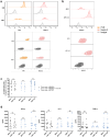
(a) Flow cytometry staining of PVR and nectin-2 (N2) in U266 and ARP-1 cells. Gray = Isotype; orange = PVR-expressing cells; red = nectin-2-expressing cells. (b) The ARP-1 cell line was lentivirally transduced with a viral vector containing the N2 gene. Positive N2 cells were sorted by flow cytometry to obtain a 100% N2-positive ARP-1 population gray = unstained cells; red = nectin-2-expressing cells. (c) Cytotoxicity results after co-culture of the MM cell lines ARP-1 and ARP-1-N2 with ARI0002h vs. ARI0002h + TIGIT-AB vs. ARITIGIT. Each dot represents a T cell donor; 3 repeats were performed for each condition in each donor. The number of T-cell donors was: (1) ARP1 + ARI0002h vs ARP1-N2 + ARI0002h: T cells from n = 10 healthy donors; (2) ARP1 + ARI0002h + TIGITAb vs ARP1-N2 + ARI0002h + TIGITAb n = 6 healthy donors; (3) ARP1 + ARITIGIT vs ARP1-N2 + ARITIGIT n = 6 healthy donors. Paired students’ t-test and the Wilcoxon ranked sign test were applied according to normality adherence. (d) Pro-inflammatory cytokine production of IFN-γ, IL-2 and TNF-α after 24 hours of co-culture. Each dot represents a T cell donor; 3 repeats were performed for each condition in each donor. The number of T-cell donors was: (1) ARP1 + ARI0002h vs ARP1-N2 + ARI0002h: T cells from n = 10 healthy donors; (2) ARP1 + ARI0002h + TIGITAb vs ARP1-N2 + ARI0002h + TIGITAb n = 6 healthy donors; (3) ARP1 + ARITIGIT vs ARP1-N2 + ARITIGIT n = 6 healthy donors. Paired students’ t-test and the Wilcoxon ranked sign test were applied according to normality adherence.
Figure 5.
Figure 5.
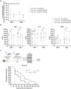
(a) Cytotoxicity results after co-culture of the MM cell lines ARP-1 and U266 with either T52 gRNA-ARI0002h or T45 gRNA-ARI0002h. Results obtained were normalized to the WT-ARI0002h group. Each dot represents a T cell donor; 3 repeats were performed for each condition in each donor. The number of T-cell donors was: (1) tumor cells + T52 gRNA-ARI0002h: n = 5 healthy donors; (2) tumor cells + T45 gRNA-ARI0002h: n = 4 healthy donors. (b) Pro-inflammatory cytokine production of IFN-γ, IL-2 and TNF-α after 24 hours of co-culture. Each dot represents a T cell donor; 3 repeats were performed for each condition in each donor. The number of T-cell donors was: (1) tumor cells + WT-ARI0002h: n = 5 healthy donors (2) tumor cells + T52 gRNA-ARI0002h: n = 5 healthy donors; (2) tumor cells + T45 gRNA-ARI0002h: n = 4 healthy donors. (c) In vivo results: mice were administered 1.5x10⁶ MM ARP-1 cells on day 0, 24 h after radiation with 2 Gy. When disease luminescence was detectable, mice were injected with 1.5x10⁶ of untransduced (UT) vs. WT-ARI0002h vs. TIGIT KO-ARI0002h. The number of mice in the experiment was: UT only n = 1, WT n = 9 and TIGIT KO n = 18. Mice were followed weekly using bioluminescence and sacrifice was performed when prespecified criteria were met. To analyze survival the log-rank test was applied and survival curves were obtained using the Kaplan-Meier method.

Similar articles

References

    1. Munshi NC, Anderson LD, Shah N, Madduri D, Berdeja J, Lonial S, Raje N, Lin Y, Siegel D, Oriol A, et al. Idecabtagene Vicleucel in relapsed and refractory multiple myeloma. N Engl J Med. 2021;384(8):705–16. doi: 10.1056/NEJMoa2024850. - DOI - PubMed
    1. Rodriguez-Otero P, Ailawadhi S, Arnulf B, Patel K, Cavo M, Nooka AK, Manier S, Callander N, Costa LJ, Vij R, et al. Ide-cel or standard regimens in relapsed and refractory multiple myeloma. N Engl J Med [Internet]. 2023;388(11):1002–1014. doi: 10.1056/NEJMoa2213614. - DOI - PubMed
    1. Martin T, Usmani SZ, Berdeja JG, Agha M, Cohen AD, Hari P, Avigan D, Deol A, Htut M, Lesokhin A, et al. Ciltacabtagene Autoleucel, an anti–b-cell maturation antigen chimeric antigen receptor T-Cell therapy, for relapsed/refractory multiple myeloma: CARTITUDE-1 2-year follow-up. J Clin Oncol. 2023. Feb 20;41(6):1265–1274. doi: 10.1200/JCO.22.00842. - DOI - PMC - PubMed
    1. San-Miguel J, Dhakal B, Yong K, Spencer A, Anguille S, Mateos M-V, Fernández de Larrea C, Martínez-López J, Moreau P, Touzeau C, et al. Cilta-cel or standard Care in Lenalidomide-refractory multiple myeloma. N Engl J Med [Internet]. 2023;389(4):335–347. doi: 10.1056/NEJMoa2303379. - DOI - PubMed
    1. Oliver-Caldés A, González-Calle V, Cabañas V, Español-Rego M, Rodríguez-Otero P, Reguera JL, López-Corral L, Martin-Antonio B, Zabaleta A, Inogés S, et al. Fractionated initial infusion and booster dose of ARI0002h, a humanised, BCMA-directed CAR T-cell therapy, for patients with relapsed or refractory multiple myeloma (CARTBCMA-HCB-01): a single-arm, multicentre, academic pilot study. Lancet Oncol [Internet]. 2023;24(8):913–924. 10.1016/S1470-2045(23)00222-X. - DOI - PubMed

LinkOut - more resources