. 2025 Sep;14(25):e2501190.
doi: 10.1002/adhm.202501190. Epub 2025 Jul 14.

Lymphoid and CXCR4 Cell Targeted Lipid Nanoparticles Facilitate HIV-1 Proviral DNA Excision

Affiliations

Lymphoid and CXCR4 Cell Targeted Lipid Nanoparticles Facilitate HIV-1 Proviral DNA Excision

Sudipta Panja et al. Adv Healthc Mater. 2025 Sep.

Abstract

Advancements in antiretroviral therapy (ART) enable those living with the human immunodeficiency virus type one (HIV-1) to lead longer, healthier lives free from disease comorbidities. However, lifelong ART poses challenges. These include social stigma, medication costs, drug accessibility, mental health, and drug-related toxicities. Moreover, ART does not eliminate latent HIV-1 DNA. Viral persistence in tissue and cell reservoirs results in viral rebound after ART interruption. New strategies are required to achieve a functional HIV-1 cure. To excise latent HIV-1, C-X-C motif chemokine receptor 4 (CXCR4) ligand-decorated lymphoid tissue-targeting lipid nanoparticles (LNPs) for CRISPR-Cas9/gRNA delivery are developed. These LNPs enhance mRNA translation and demonstrate CXCR4-mediated improved uptake to eliminate HIV-1 DNA in infected CD4+ T cells. LNPs also facilitate targeted drug delivery, achieving HIV-1 DNA excision in ART-treated, infected humanized mice. This study emphasizes the potential of tissue and cell-targeted LNPs for effective HIV-1 DNA excision.

Keywords: CRISPR; HIV‐1 infection; excision of HIV‐1 DNA; lipid nanoparticles; targeted mRNA delivery.


Conflict of interest statement

The authors declare that Dr. Howard Gendelman is co‐founder of Exavir Therapeutics, Inc. The biotechnology company is developing ultra‐long‐acting drugs. The drugs in development are not linked to those created in the current report. All other authors declare no competing interests.

Figures

Scheme 1
Scheme 1
Schematic illustration of the synthesis of DSPE‐PEG‐CycPep via an activated ester‐amine coupling reaction.
Figure 1
Figure 1
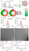
LNP design and characterization. A) Schematic representation of LNP formulation by microfluidic mixing and subsequent purification. Created with BioRender. B) Donut charts showing the molar composition of the lipids used to formulate C‐LNP and T‐LNP. C,D) Size distribution of C‐LNP and T‐LNP determined by dynamic light scattering (DLS). E,F) Cryo‐TEM images of LNPs. G) mRNA encapsulation efficiency of LNPs, evaluated using the RiboGreen assay, indicates >97% encapsulation for both formulations. H,I) The long‐term stability of LNPs was assessed through size and polydispersity index (PDI) measurements (n = 3), showing no significant changes.
Figure 2
Figure 2
Tissue selective mRNA translation of LNPs. A,B) Tissue‐selective mRNA translation efficiency of LNPs in BALB/c and humanized mice. C‐LNP exhibits liver‐selective mRNA translation, while T‐LNP shows spleen‐selective mRNA translation. C) Spleen‐to‐liver luminescence ratio in humanized mice (n = 3), highlighting the spleen tissue selectivity of T‐LNP. The mRNA translation efficiency was evaluated after tail vein administration of LNPs at 0.5 mg kg−1 mRNA dose. Luminescence images were acquired 6 h following administration using an in vivo imaging system (IVIS). Data are presented as means ± SD (n = 3). *** p < 0.001.
Figure 3
Figure 3
CXCR4‐dependent uptake and mRNA translation. A) Schematic representation of latent HIV‐1‐infected cell lines (lymphocytic JLat 8.4, J1.1, and promonocytic U1) and their respective CXCR4 expression. Created with BioRender. B) Quantification of CXCR4 expression by flow cytometry. J1.1 cells show the highest CXCR4 expression, followed by JLat 8.4 and U1 cells. C) Luciferase mRNA translation efficacy of LNPs across different cell lines. The mRNA translation efficiency of these LNPs was assessed 48 h post‐LNP treatment (at 1 µg/million cells mRNA dose) using the luciferase assay and presented as relative luminescence units (RLU). D,E) Concentration‐dependent CXCR4‐receptor inhibition by AMD070 and its impact on mRNA translation efficiency of T‐LNP and C‐LNP across various cell lines. Data are presented as means ± SD (n = 3). F) Effect of AMD070 pretreatment on the cellular uptake of Cy5.5‐labeled T‐LNPs in J1.1 cells. Cells were pretreated with AMD070 for 30 min, followed by treatment with Cy5.5‐labeled T‐LNPs. After 4 h, cells were co‐stained with DAPI (blue) and phalloidin (green) and visualized using confocal microscopy.
Figure 4
Figure 4
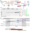
HIV‐1 proviral DNA excision efficacy of CRISPR‐Cas9 LNPs. A,B) Schematic of the CRISPR‐Cas9 gene‐editing process and multiple doasing. Created with BioRender. C) PCR gel electrophoresis image of DNA extracted from JLat 8.4 cells treated with LNPs (1 µg/10⁶ cells) formulated with varying gRNA/m1Ψ‐mCas9 molar ratios. Red arrowheads denote the proviral DNA amplicon (3 kb) and the excised DNA amplicon (428 bp). D) Quantifying HIV‐1 DNA excision efficacy of LNPs formulated with different gRNA/m1Ψ‐Cas9 molar ratios, analyzed using ImageJ and densitometric analysis. E–H) The comparison and quantification of HIV‐1 DNA excision efficacy between T‐LNP and C‐LNP in lymphocytic (E, F) JLat 8.4 and (G, H) J1.1 cell lines. I) Effect of AMD070 pretreatment on the HIV‐1 DNA excision efficacy of T‐LNPs in JLat 8.4 and J1.1 cell lines. J) The differences in excision efficacy following AMD070 treatment were quantified using ImageJ and densitometric analysis. K,L) PCR gel electrophoresis image and quantification of HIV‐1 DNA excision efficacy following multiple doses of LNP treatment in the JLat 8.4 cell line. Data are presented as means ± SD (n = 3). ** p < 0.01 and **** p < 0.0001.
Figure 5
Figure 5
HIV‐1 proviral DNA excision efficacy in lymphoblasts. A) Timeline for elutriation, cell separation, culture, HIV‐1 infection, ART and LNP treatment, and DNA extraction of PBLs collected from healthy, HIV‐1 seronegative donors. Created with BioRender. IL‐2 and phytohemagglutinin‐treated PBLs (lymphoblasts) were infected with HIV‐1NL4‐3 at 0.001 MOI for 24 h and subsequently subjected to ART for 48 h. After ART treatment, PBLs were treated with LNPs at an mRNA dose of 8 µg/million cells. PBLs were harvested 72 h after treatment for DNA extraction and PCR. B) PCR gel electrophoresis image shows near‐complete excision of HIV‐1 proviral DNA using T‐LNP. The proviral (3 kb) and excised (428 bp) DNA amplicons are highlighted by red arrowheads. C) The excision efficacy of T‐LNP was quantified using ImageJ and densitometric analysis. D) Excised amplicons were sequenced and confirmed as CRISPR edits by Sanger sequencing. Multiple sequence alignment was performed to align the amplicon sequences with the HIV‐1NL4‐3 reference genome (FWD = forward strand, REV = reverse strand). Data are presented as means ± SD (n = 3). **** p < 0.0001.
Figure 6
Figure 6
Excision of HIV‐1 proviral DNA in hu mice. A) The study timeline includes blood collection for immune cell profiling, HIV infection, ART and LNP treatments; the study endpoint involves the sacrifice of hu mice (n = 3) and the blood and tissue collection. B) The plasma HIV‐1 RNA copies at 3‐, 9‐ and 11‐weeks post‐infection (WPI). C,D) The levels of CD45+ and CD4+ cells in blood were analyzed by flow cytometry. E) The schematic representation of the HIV‐1NL4‐3 genome highlights the Ψ and env probes together with TatDE binding sites. F) The representative illustration of intact proviral DNA assay (IPDA) 2D duplexed droplet digital PCR plot. Q1(blue), Q2(orange), Q3 (gray) and Q4 (green) quadrants represent Ψ‐single‐positive, Ψ and env double‐positive, double‐negative, and env single‐positive events, respectively. The HIV‐1 proviral DNA excision efficacy was analyzed by comparing the number of Ψ‐positive and env‐positive events. G–I) The HIV‐1 DNA excision efficacy of LNPs in spleen, blood, and lung, analyzed by IPDA assay. T‐LNPs achieved ≈60% HIV DNA excision efficacy in the spleen and blood, while C‐LNPs showed ≤5% excision efficacy. Comparable efficacy was observed in the lungs, but lower than the spleen and blood. Data are presented as means ± SD (n = 3). Figures A and E were created with BioRender.
Figure 7
Figure 7
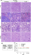
Toxicology evaluation. A) Hematoxylin and eosin staining of liver, spleen, lungs, and kidney tissues from each treatment group imaged at 20× magnification. B) The AST and ALT ratios along the various treatment groups. C) Mice body weight over the treatment period, presented by weeks after infection. No significant changes in body weight were observed, indicating the safety of LNP treatment. Data are presented as means ± SD (n = 3).

References

    1. WHO, HIV Factsheets , https://www.who.int/news‐room/fact‐sheets/detail/hiv‐aids, accessed.
    1. Koethe J. R., Lagathu C., Lake J. E., Domingo P., Calmy A., Falutz J., Brown T. T., Capeau J., Nat. Rev. Dis. Primers 2020, 6, 48. - PubMed
    1. Chun T.‐W., Finzi D., Margolick J., Chadwick K., Schwartz D., Siliciano R. F., Nat. Med. 1995, 1, 1284. - PubMed
    1. Chun T.‐W., Carruth L., Finzi D., Shen X., DiGiuseppe J. A., Taylor H., Hermankova M., Chadwick K., Margolick J., Quinn T. C., Kuo Y.‐H., Brookmeyer R., Zeiger M. A., Barditch‐Crovo P., Siliciano R. F., Nature 1997, 387, 183. - PubMed
    1. Neumann A. U., Tubiana R., Calvez V., Robert C., Li T.‐S., Agut H., Autran B., Katlama C., AIDS 1999, 13, 677. - PubMed