. 2025 Jun;15(6):3041-3058.
doi: 10.1016/j.apsb.2025.04.017. Epub 2025 Apr 22.

SRSF7 promotes pulmonary fibrosis through regulating PKM alternative splicing in lung fibroblasts

Affiliations

SRSF7 promotes pulmonary fibrosis through regulating PKM alternative splicing in lung fibroblasts

Tongzhu Jin et al. Acta Pharm Sin B. 2025 Jun.

Abstract

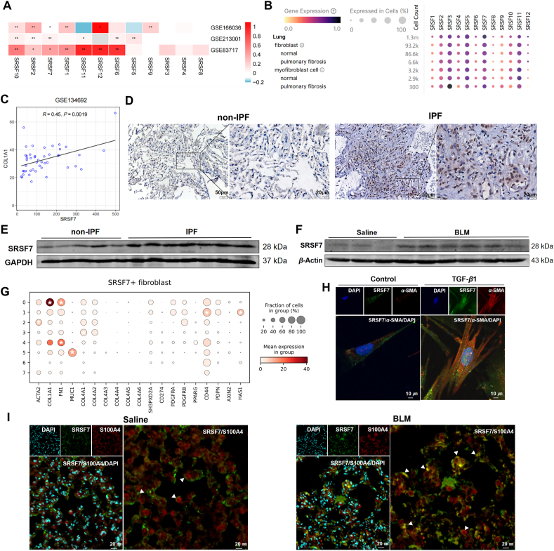
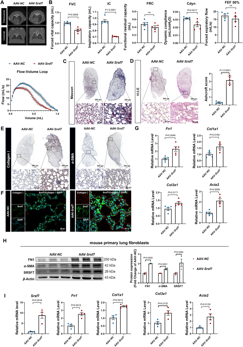
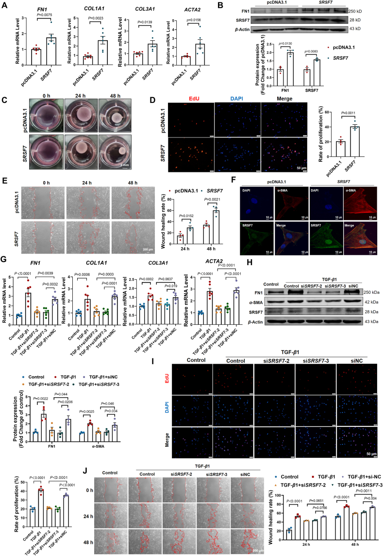
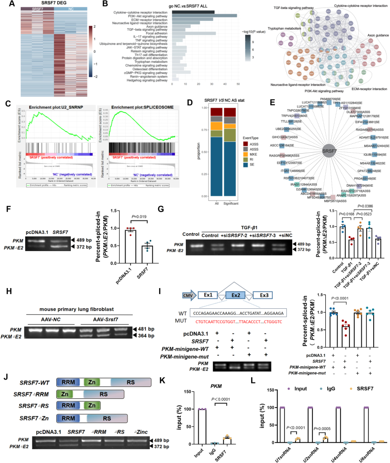
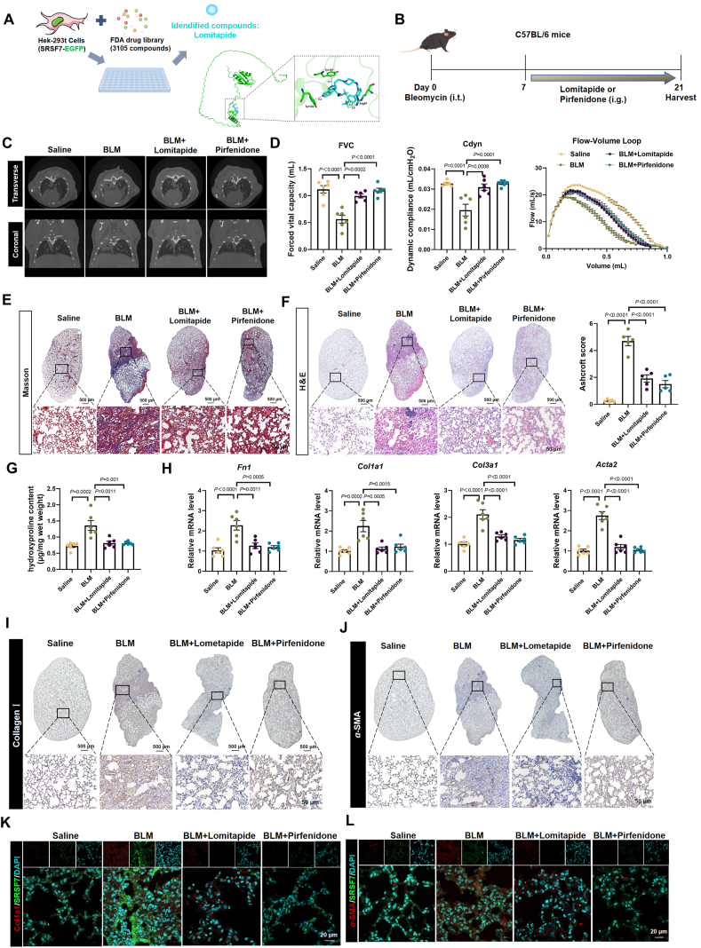
Idiopathic pulmonary fibrosis (IPF), a chronic interstitial lung disease, is characterized by aberrant wound healing, excessive scarring and the formation of myofibroblastic foci. Although the role of alternative splicing (AS) in the pathogenesis of organ fibrosis has garnered increasing attention, its specific contribution to pulmonary fibrosis remains incompletely understood. In this study, we identified an up-regulation of serine/arginine-rich splicing factor 7 (SRSF7) in lung fibroblasts derived from IPF patients and a bleomycin (BLM)-induced mouse model, and further characterized its functional role in both human fetal lung fibroblasts and mice. We demonstrated that enhanced expression of Srsf7 in mice spontaneously induced alveolar collagen accumulation. Mechanistically, we investigated alternative splicing events and revealed that SRSF7 modulates the alternative splicing of pyruvate kinase (PKM), leading to metabolic dysregulation and fibroblast activation. In vivo studies showed that fibroblast-specific knockout of Srsf7 in conditional knockout mice conferred resistance to bleomycin-induced pulmonary fibrosis. Importantly, through drug screening, we identified lomitapide as a novel modulator of SRSF7, which effectively mitigated experimental pulmonary fibrosis. Collectively, our findings elucidate a molecular pathway by which SRSF7 drives fibroblast metabolic dysregulation and propose a potential therapeutic strategy for pulmonary fibrosis.

Keywords: Alternative splicing; Drug screening; Fibroblasts; IPF; Metabolism; PKM; SRSF7; Splicing factor.


Conflict of interest statement

The authors declared no conflict of interest.

Figures

Image 1
Graphical abstract
Figure 1
Figure 1
Increased expression of SRSF7 in lung fibroblasts during pulmonary fibrosis. (A) Heatmap of mRNA expression levels of serine/arginine-rich splicing factors in lung samples from GSE166036, GSE213001 and GSE83717. The color bars represent the range of log2FC values for each gene, with higher log2FC values shown in red and lower values in blue. ∗P < 0.05, ∗∗P < 0.01. (B) Single-cell RNA sequencing of annotated human lung fibrosis populations generated from Chan-Zuckerberg CELL by GENE Discover online database depicting relative expression of SRSF gene. Circle color denotes mean gene expression within each fibroblast subtype while circle size represents the proportion of each cell population expressing the indicated gene. (C) The Pearson’s correlation analysis between the expression of COL1A1 and SRSF7 in the advanced IPF samples from GSE134692 dataset. The X-axis shows the FPKM-normalized expression values of the SRSF7 gene. The Y-axis shows the expression level of COL1A1. Pearson correlation (r) and P-value (P) are shown. (D) The IHC staining of SRSF7 in non-IPF and IPF patients. Non-IPF: n = 6, IPF: n = 8. (E) The protein expression of SRSF7 in lung tissues. Non-IPF: n = 5, IPF: n = 7. (F) The protein expression of SRSF7 in BLM treated mice. Saline: n = 3, BLM: n = 6. (G) In GSE159354, marker genes were identified using the rank_genes_groups function in Scanpy, with the wilcoxon test applied. Genes with a log2FC ≥ 4 and P < 0.05 were considered markers and were marked with an asterisk. (H) Dual immunofluorescence in TGF-β1 treated MRC-5 cells. n = 3, scale bar = 10 μm. (I) Dual immunofluorescence of S100A4 (red) and SRSF7 (green) in frozen lung sections. n = 3, scale bar = 20 μm.
Figure 2
Figure 2
Overexpression of SRSF7 led to pulmonary function decline. (A) micro-CT was used to observe the fibrosis of mouse lung; n = 5. (B) FVC, IC, FRC, Cdyn, FEF 50% and Flow-volume loop were detected in mice treated with AAV-NC and AAV-Srsf7; n = 5. (C) Masson staining was used to evaluate the content of collagen in lung tissues; n = 4, scale bar = 500 μm. (D) The alveoli structure detected by H&E staining and the degree of interstitial lung fibrosis were determined by using a predetermined numerical scale of 0–8, based on the Ashcroft scoring method; n = 5, scale bar = 500 μm. (E, F) Collagen I and α-SMA were stained by IHC (n = 3, scale bar = 500 μm) and immunofluorescence staining (n = 4, scale bar = 20 μm). (G) The mRNA expression of Fn1, Col1α1, Col3α1 and Acta2 in mice treated with AAV-NC and AAV-Srsf7; n = 5. (H) The protein expression of FN1, α-SMA and SRSF7 in the lung fibroblasts of mice. (I) The mRNA expression of fibrosis markers in mice treated with AAV-NC and AAV-Srsf7; n = 4. Data are presented as mean ± SEM; ns, no significance.
Figure 3
Figure 3
SRSF7 promotes fibroblast activation and fibrogenesis. (A) The mRNA expression of fibrosis related genes after MRC-5 cells transfected with SRSF7; n = 6. (B) Western blot demonstrated increased expression level of Fn1 in SRSF7 transfected MRC-5 cells; n = 3. (C) Relative quantification of the collagen contraction area after transfected with SRSF7; n = 3. (D, E) EdU assay (scale bar = 50 μm, n = 4) and wound healing experiment (scale bar = 200 μm, n = 4) indicated that transfected with SRSF7 promoted the proliferation and migration of MRC-5. (F) Overexpression of SRSF7 promoted the fibroblast-myofibroblast transformation of MRC-5 cells; n = 3, scale bar = 10 μm. (G) qRT-PCR analysis demonstrating the relative expression of FN1, COL1A1, COL3A1 and ACTA2 in TGF-β1-induced MRC-5 cells transfected with or without si-SRSF7; n = 6. (H) Western blot showed that the si-SRSF7 abrogated the upregulation of fibrotic proteins in MRC-5 cells induced by TGF-β1; n = 4. (I, J) EdU (scale bar = 50 μm, n = 4) and wound healing assays (scale bar = 200 μm, n = 3) indicated that si-SRSF7 inhibited the proliferation and migration of MRC-5 cells. Data are presented as mean ± SEM.
Figure 4
Figure 4
SRSF7 is able to bind to and regulate the alternative splicing of PKM. (A) Heatmap illustrating differentially expressed genes. Red represents up-regulated, and blue represents down-regulated. In the heatmap, red indicates high gene expression, while blue represents low gene expression. The color bars display the scale of values for each gene, ranging from −2 to 2 as shown on the right. (B) GO enrichment analysis regulated by overexpression of SRSF7. (C) GSEA showed that U2 SnRNP and spliceosome related genes were significantly enriched after overexpression of SRSF7. (D) The differentially alternative splicing genes regulated by SRSF7 were mainly enriched in exon skipped events. (E) Prediction of SRSF7 regulated alternative splicing genes overlap in MRC-5 and IPF patients. (F) SRSF7 enhanced the alternative splicing of PKM exon 2; n = 4. (G) Silencing SRSF7 can reduce the skipping of PKM exon 2 induced by TGF-β1; n = 4. (H) Lung fibroblasts were isolated from mice to detect the alternative splicing events of PKM. (I) Minigene reporters of PKM were introduced into SRSF7-overexpression cells; n = 6. (J) Deficiency of the SRSF7 function domain on alternative splicing of PKM exon 2. (K) RIP experiments were performed to detect the binding relationship between SRSF7 and PKM; n = 3. (L) The effect of SRSF7 on the ability of binding with snRNAs was investigated by the RIP experiment; n = 3. Data are presented as mean ± SEM.
Figure 5
Figure 5
PKME2 isoform promotes fibroblasts activation. (A) The mRNA expression of fibrosis related genes; n = 6. (B) Western blot suggested that PKM isoforms promote the expression of FN1 and α-SMA; n = 5. (C, D) EdU (scale bar = 50 μm, n = 4) and wound healing assays (scale bar = 200 μm, n = 3) indicated that PKM isoforms can promote the cell proliferation and migration. (E) Relative quantification of the collagen contraction area after overexpression of PKM isoforms; n = 3. (F) Overexpression of PKME2 increased the pruvate activity and lactic acid content compared with PKM. (G, H) MRC-5 cells were seeded in Seahorse XF-96 cell culture microplates. The cells were transfected with PKM and PKME2, followed by sequential treatments with oligomycin (Oligo) and FCCP. Real-time ECAR and OCR were recorded. Data are presented as mean ± SEM.
Figure 6
Figure 6
Silencing SRSF7 alleviates fibrogenesis by inhibiting the expression of PKME2 isoform. (A) The mRNA expression of FN1, COL1A1, COL3A1 and ACTA2; n = 4. (B) Relative quantification of the collagen contraction area in MRC-5 cells; n = 3. (C) Immunofluorescence showed the fibroblasts transformation in MRC-5; n = 3, scale bar = 20 μm. (D, E) EdU (scale bar = 50 μm, n = 3) and wound healing assays (scale bar = 200 μm, n = 3) indicated that overexpression of PKME2 promoted the cell proliferation and migration reduced by siSRSF7. (F, G) Real-time ECAR and OCR were recorded in the MRC-5 transfected with siSRSF7 and PKME2 after treatment with TGF-β1. (H) The lactic acid content of MRC-5 cells, n = 6. Data are presented as mean ± SEM.
Figure 7
Figure 7
Srsf7 deficiency attenuates BLM-induced experimental lung fibrosis in mice. (A) Diagram of the animal experimental model of Srsf7-cKO. (B) Representative micro-CT images showed the lung fibrosis of Srsf7-cKO mice treated with bleomycin for 21 days; n = 4. (C, D) FRC, FVC, IC, FEF50%, Cdyn and F–V Loop were detected in Srsf7fl/fl or Srsf7-cKO mice treated with saline or bleomycin for 21 days; n = 6. (E) Masson and (F) H&E staining were used to evaluate the content of collagen in lung tissues; n = 6, scale bar = 500 μm. (G) The hydroxyproline content detection in lung tissues was confirmed by hydroxyproline detection kit; n = 6. (H) qRT-PCR was used to determine the mRNA level of Srsf7, Fn1, Acta2, Col1a1 and Col3a1; n = 6. (I, J) IHC (n = 3, scale bar = 500 μm) and (K, L) immunofluorescence staining (n = 4, scale bar = 20 μm) were used to evaluate the expression of Collagen Ⅰ and α-SMA in lung tissues. Data are presented as mean ± SEM.
Figure 8
Figure 8
Lomitapide inhibits SRSF7 expression to improve lung fibrosis and lung function in mice. (A) Chemical screen to systematically identify inhibitors of SRSF7 and prediction of Drug–Protein Interaction Networks from the Integration of SRSF7 Sequences and Lomitapide Chemical Structures (right panel). (B) Experimental protocol of lomitapide in BLM treated mice. (C) Representative micro-CT images showed the lung fibrosis of lomitapide treated mice; n = 4. (D) FVC, Cdyn and F–V Loop were detected in mice treated with lomitapide or pirfenidone; n = 6. (E) Masson and (F) H&E staining were used to evaluate the content of collagen in lung tissues; n = 5, scale bar = 500 μm. (G) The hyroxyproline content detection in lung tissues was confirmed by hydroxyproline detection kit; n = 6. (H) qRT-PCR was used to determine the mRNA level of Fn1, Acta2, Col1a1 and Col3a1 in lomitapide or pirfenidone treated mice; n = 6. (I, J) IHC (n = 4, scale bar = 500 μm) and (K, L) immunofluorescence staining (n = 3, scale bar = 20 μm) were used to evaluate the expression of Collagen Ⅰ and α-SMA in lung tissues. Data are presented as mean ± SEM.

References

    1. Cui H., Xie N., Banerjee S., Ge J., Jiang D., Dey T., et al. Lung myofibroblasts promote macrophage profibrotic activity through lactate-induced histone lactylation. Am J Respir Cell Mol Biol. 2021;64:115–125. - PMC - PubMed
    1. Raghu G., Collard H.R., Egan J.J., Martinez F.J., Behr J., Brown K.K., et al. An official ATS/ERS/JRS/ALAT statement: idiopathic pulmonary fibrosis: evidence-based guidelines for diagnosis and management. Am J Respir Crit Care Med. 2011;183:788–824. - PMC - PubMed
    1. Richeldi L., Collard H.R., Jones M.G. Idiopathic pulmonary fibrosis. Lancet. 2017;389:1941–1952. - PubMed
    1. Zhao L., Zhu Y., Tao H., Chen X., Yin F., Zhang Y., et al. Ailanthone ameliorates pulmonary fibrosis by suppressing JUN-dependent MEOX1 activation. Acta Pharm Sin B. 2024;14:3543–3560. - PMC - PubMed
    1. Fortier S.M., Walker N.M., Penke L.R., Baas J.D., Shen Q., Speth J.M., et al. MAPK phosphatase 1 inhibition of p38alpha within lung myofibroblasts is essential for spontaneous fibrosis resolution. J Clin Investig. 2024;134 - PMC - PubMed

LinkOut - more resources