. 2025 Jun;15(6):3149-3162.
doi: 10.1016/j.apsb.2025.04.001. Epub 2025 Apr 4.

Psychological stress-activated NR3C1/NUPR1 axis promotes ovarian tumor metastasis

Affiliations

Psychological stress-activated NR3C1/NUPR1 axis promotes ovarian tumor metastasis

Bin Liu et al. Acta Pharm Sin B. 2025 Jun.

Abstract

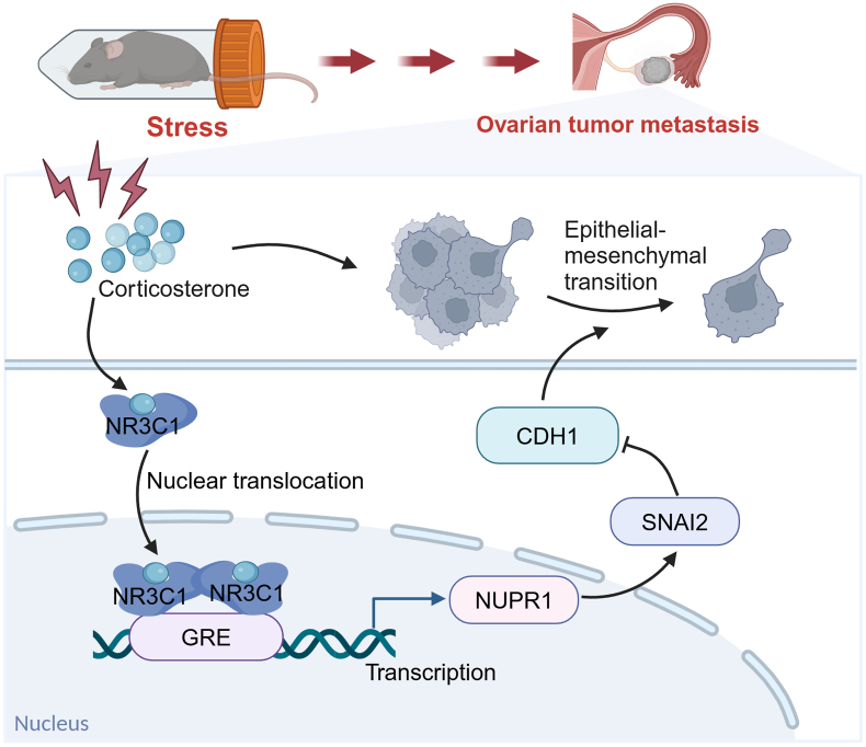
Ovarian tumor (OT) is the most lethal form of gynecologic malignancy, with minimal improvements in patient outcomes over the past several decades. Metastasis is the leading cause of ovarian cancer-related deaths, yet the underlying mechanisms remain poorly understood. Psychological stress is known to activate the glucocorticoid receptor (NR3C1), a factor associated with poor prognosis in OT patients. However, the precise mechanisms linking NR3C1 signaling and metastasis have yet to be fully elucidated. In this study, we demonstrate that chronic restraint stress accelerates epithelial-mesenchymal transition (EMT) and metastasis in OT through an NR3C1-dependent mechanism involving nuclear protein 1 (NUPR1). Mechanistically, NR3C1 directly regulates the transcription of NUPR1, which in turn increases the expression of snail family transcriptional repressor 2 (SNAI2), a key driver of EMT. Clinically, elevated NR3C1 positively correlates with NUPR1 expression in OT patients, and both are positively associated with poorer prognosis. Overall, our study identified the NR3C1/NUPR1 axis as a critical regulatory pathway in psychological stress-induced OT metastasis, suggesting a potential therapeutic target for intervention in OT metastasis.

Keywords: EMT; NR3C1; NUPR1; Ovarian tumor; Prognostic biomarker; Psychological stress; SNAI2; Therapeutic target.


Conflict of interest statement

The authors declare no competing interests.

Figures

Image 1
Graphical abstract
Figure 1
Figure 1
Psychological stress-evoked OT metastasis is mediated by NR3C1 activation. (A) Time course of the experimental procedure. Mice were orthotopically injected with ID8-Luc cells, and then exposed to restraint stress daily for 6 weeks. (B–E) The OT marker CA125, tumor photograph, tumor weight, and tumor volume were measured on the 7th week (n = 6). (F, G) The OT metastasis in vivo was imaged by a living imaging system, and the luminescence intensity was quantified (n = 3). Metastatic foci at (H, I) intestine, (J, K) peritoneum and (L, M) diaphragm in mice were measured and quantified (n = 5–6). (N) The corticosterone level from the serum of mice was detected by LC/MS (n = 6). (O, P) The activation of NR3C1 in OT tissue was measured by IHC (n = 3). (Q) Representative fluorescence imaging of OT metastasis in mice treated with Stress, CORT, and Stress + Ru486 (n = 3). All data represent mean ± SD. ∗P < 0.05, ∗∗P < 0.01, ∗∗∗P < 0.001, by independent-samples t test.
Figure 2
Figure 2
NR3C1 activation contributes to the invasion and migration of OT in vitro. (A) NR3C1 expression of SKOV3, HO8910, and A2780 cells was detected by Western blot (n = 3). (B) Cell growth of SKOV3 treated with cortisol for 48 and 96 h was detected by MTT (n = 3). (C) Cellular morphology of SKOV3 and HO8910 cells treated with cortisol or Ru486 for 48 h was observed by microscope (n = 3). (D) Cell migration of SKOV3 cells treated with cortisol or Ru486 for 48 and 96 h were detected by wound healing (n = 3). (E, F) Invasion and migration of SKOV3 cells treated with cortisol or Ru486 for 24 h were detected by the Transwell method (n = 3). (G) The effect of NR3C1 activation in SKOV3 cells treated with cortisol or siNR3C1 was observed by immunofluorescence (n = 3). (H, I) Invasion and migration of SKOV3 cells treated with CORT or siNR3C1 for 24 h were detected by the Transwell method (n = 3). All data represent mean ± SD. ∗P < 0.05, ∗∗P < 0.01, ∗∗∗P < 0.001, by one-way ANOVA with Tukey.
Figure 3
Figure 3
NUPR1 expression is highly correlated with GC-induced NR3C1 activation. (A) The KEGG pathway enrichment analysis of differential genes in SKOV3 cells treated with or without cortisol (n = 3). (B) Expression profile of differential genes related to pathways in cancer, transcriptional mis-regulation in cancer, and focal adhesion in SKOV3 cells (n = 3). (C) The biological process enrichment analysis of differential genes in SKOV3 cells treated with or without cortisol (n = 3). (D) The GO chord indicates overlapped the differential genes across the biological process. (E–G) The mRNA and protein levels of NUPR1 and SNAI2 in SKOV3 cells treated with siNR3C1 for 48 h were analyzed (n = 3). (H) Correlation of NR3C1 mRNA level and NUPR1 mRNA level in human ovarian cancer samples (TCGA PanCancer Atlas Studies, cBioportal). mRNA level was batch normalized from Illumina HiSeq_RNASeqV2 and shown as log2 (RSEM + 1). R2 = 0.12, Spearman correlation. All data represent mean ± SD. ∗∗P < 0.01, by independent-samples t-test (for E and G).
Figure 4
Figure 4
NUPR1 is required for OT metastasis and NR3C1-dependent transcriptional regulated by GC. (A–C) The silencing effect of siNUPR1 in SKOV3 cells was detected by qRT-PCR and Western blot (n = 4). (D–F) The mRNA and protein level of SNAI2 and CDH1 in SKOV3 cells treated with siNUPR1 for 24 h were detected (n = 3). (G, H) The cell invasion of SKOV3 cells treated with CORT/siNUPR1 for 24 h was detected by the Transwell method (n = 3). (I) Schematic map of putative GREs within the 2000 bp region of human NUPR1 promoter. (J, K) Luciferase activities of NUPR1 were determined in NR3C1 plasmids or cortisol-treated HEK 293 cells (n = 3). (L) The enrichment of NR3C1 to the GRE site at NUPR1 promoter region in HEK293 cells treated with cortisol was analyzed by ChIP assay (n = 3). All data represent mean ± SD. ∗P < 0.05, ∗∗P < 0.01, ∗∗∗P < 0.001, by one-way ANOVA with Tukey.
Figure 5
Figure 5
NR3C1 and NUPR1 expression are correlated to the poor prognosis of OT of patients. KM survival analysis for the relationship between survival time, NR3C1 and NUPR1 signatures in OT was performed by using the online tool (http://kmplot.com/analysis/). (A–C) Overall survival (n = 1656) and progression-free survival (n = 1435) in OT patients. (D) The representative Magnetic Resonance Imaging images in OT (n = 75), ovarian endometrial cysts (OC, n = 9) and the normal tissues (Normal, n = 5) of patients. The green circle indicates the normal ovary of the patient with uterine fibroids; the red circle indicates the OT tissue of I/II/III stage patients; the red arrow indicates the ascites of III stage OT patients; and the yellow circle indicates ovarian cyst tissue of patients with ovarian cyst. (E) Representative IHC staining image and (F) quantification of NR3C1 expression in OT, OC, and normal tissue of patients. “T” indicates the tumor tissue and red arrow indicates NR3C1 positive staining. (G) The level of NR3C1 expression in low grade and high grade of OT tissue. (H) The level of NR3C1 expression in II/III/IV stage and I stage of OT tissue. (I, J) Representative IHC staining image and the quantification of NUPR1 in OT, OC, and normal patients. The “T” indicates tumor tissue and the red arrow indicates NUPR1 positive staining. ∗P < 0.05, ∗∗P < 0.01, ∗∗∗P < 0.001.
Figure 6
Figure 6
The illustration shows that psychological stress-induced glucocorticoid receptor activation promotes NUPR1 transcription, thereby evoking ovarian tumor metastasis. Created with BioRender.com.

References

    1. Webb P.M., Jordan S.J. Epidemiology of epithelial ovarian cancer. Best Pract Res Clin Obstet Gynaecol. 2017;41:3–14. - PubMed
    1. Siegel R.L., Miller K.D., Jemal A. Cancer statistics, 2020. CA Cancer J Clin. 2020;70:7–30. - PubMed
    1. Markman M. Optimal management of recurrent ovarian cancer. Int J Gynecol Cancer. 2009;19(Suppl 2):S40–S43. - PubMed
    1. Armer J.S., Clevenger L., Davis L.Z., Cuneo M., Thaker P.H., Goodheart M.J., et al. Life stress as a risk factor for sustained anxiety and cortisol dysregulation during the first year of survivorship in ovarian cancer. Cancer. 2018;124:3401–3408. - PMC - PubMed
    1. Liu C.L., Liu L., Zhang Y., Dai X.Z., Wu H. Prevalence and its associated psychological variables of symptoms of depression and anxiety among ovarian cancer patients in China: a cross-sectional study. Health Qual Life Outcome. 2017;15:161. - PMC - PubMed

LinkOut - more resources