Review
. 2025 Jun 26:30:278-298.
doi: 10.1016/j.reth.2025.06.008. eCollection 2025 Dec.

Regenerative potential of PRP-based scaffolds in chronic wound healing: Mechanisms, advances, and therapeutic insights

Affiliations
Review

Regenerative potential of PRP-based scaffolds in chronic wound healing: Mechanisms, advances, and therapeutic insights

Farag M A Altalbawy et al. Regen Ther. .

Abstract

Introduction: Chronic wounds such as diabetic foot ulcers, venous leg ulcers, and pressure ulcers often remain trapped in the inflammatory phase due to oxidative stress, protease overactivity, and impaired cellular responses, particularly in diabetic conditions. These wounds require advanced therapeutic strategies beyond conventional care. Regenerative medicine-especially platelet-rich plasma (PRP)-based interventions-has emerged as a promising approach for enhancing wound repair.

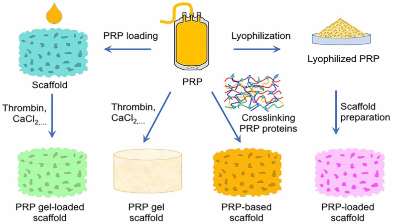
Methods: This review examines recent developments in PRP-loaded scaffolds, focusing on their biological mechanisms, structural advantages, and clinical applications. It synthesizes findings from key studies that integrate PRP with natural and synthetic biomaterials, often combined with bioactive agents like adipose-derived stem cell exosomes.

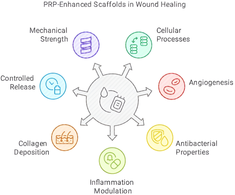
Results: PRP-containing scaffolds promote wound healing through multiple pathways: enhancing cell proliferation, migration, angiogenesis, and extracellular matrix remodeling; reducing inflammation via M2 macrophage polarization; and facilitating collagen deposition. Their antibacterial properties and controlled release of growth factors such as VEGF and TGF-β1 further support tissue regeneration. Additionally, scaffold composition improves mechanical strength, elasticity, and growth factor bioavailability. Innovations such as GelMA/SFMA hydrogels and COL/PRP-ADSC-exos composites have shown superior outcomes in preclinical models.

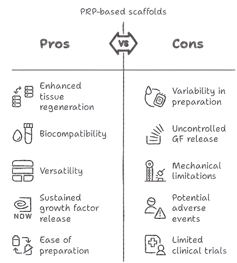
Conclusions: PRP-based scaffolds offer a multifunctional platform for chronic wound treatment by combining biological activity with structural support. Despite existing challenges such as variability in PRP preparation and limited clinical data, ongoing research and emerging technologies hold strong potential to standardize and enhance these therapies for future clinical translation.

Keywords: Angiogenesis; Chronic wound healing; Extracellular matrix remodeling; Platelet-rich plasma (PRP); Regenerative medicine; Scaffold-based therapy.


Conflict of interest statement

The authors declare that they have no known competing financial interests or personal relationships that could have appeared to influence the work reported in this paper.

Figures

Image 1
Graphical abstract
Fig. 1
Fig. 1
Phases of wound healing. The process begins with hemostasis, during which blood loss is controlled through the formation of a platelet plug and a fibrin matrix. This is followed by the inflammatory phase, where neutrophils, recruited by histamine release from mast cells, help eliminate dead cells and prevent infection. Monocytes then differentiate into macrophages, aiding in the clearance of cellular debris and neutrophils in the wound area. In the proliferative phase, multiple processes occur, including keratinocyte migration to close the wound, angiogenesis for new blood vessel formation, and fibroblast activation, which stimulates granulation tissue formation. Finally, during tissue remodeling, fibroblasts, MSCs, myofibroblasts, and blood vessels collaborate to reshape the wound site, ultimately leading to complete wound closure (Figure reprinted from Ref. [61], licensed under CC BY 4.0.).
Fig. 2
Fig. 2
Strategies for PRP incorporation in scaffolds for tissue engineering applications. PRP is either lyophilized to enhance shelf life and bioactivity before scaffold preparation (resulting in lyophilized PRP-loaded scaffolds) or directly incorporated into scaffolds through various methods. These methods include PRP gel formation via thrombin and CaCl2 activation, leading to PRP gel-loaded scaffolds, and protein cross-linking, resulting in PRP-based scaffolds with improved mechanical properties and sustained growth factor release. The use of synthetic, natural, or composite scaffolds enhances biocompatibility, mechanical stability, and regenerative efficiency, facilitating wound healing, osteogenesis, and tissue repair (Figure reprinted from Ref. [33], licensed under CC BY 4.0.).
Fig. 3
Fig. 3
Effects of PRP-Containing scaffolds on key cellular and biological processes in wound healing.
Fig. 4
Fig. 4
Ki67 Staining of Proliferating Cells. (I) Assessment of cell proliferation in wounds on day 14. Blue solid square boxes highlight regions examined at higher magnification within the full-thickness skin graft layer, while red solid square boxes indicate areas analyzed at higher magnification within the scaffold layer. High-magnification images are presented with corresponding colored frames following the overall morphological view. (II) Quantification of proliferating cells in the full-thickness skin graft layer. (III) Quantification of proliferating cells in the scaffold layer (∗P < 0.05). (Figure reprinted from Ref. [105], licensed under CC BY-NC-ND).
Fig. 5
Fig. 5
In Vivo Angiogenesis Assessment. (I) CD31 immunohistochemical staining of wound tissue sections on postoperative day 14 revealed the presence of newly formed blood vessels at the wound sites. Panels A, B, C, and D correspond to pDA-CSS@PRP, CSS@PRP, CSS, and the untreated control group, respectively. (II) Macroscopic evaluation of wound healing outcomes at day 21 post-injury, captured from the wound undersurface, illustrating differences across treatment groups. (III) Quantitative analysis of CD31-positive vessels, with statistical significance indicated (P < 0.05). (Figure reprinted from Ref. [105], licensed under CC BY-NC-ND).
Fig. 6
Fig. 6
Advantages and limitations of PRP-based scaffolds in wound healing.

References

    1. Fernández-Guarino M., Hernández-Bule M.L., Bacci S. Cellular and molecular processes in wound healing. Biomedicines. 2023;11:2526. doi: 10.3390/biomedicines11092526. - DOI - PMC - PubMed
    1. Mamun A.A., Shao C., Geng P., Wang S., Xiao J. Recent advances in molecular mechanisms of skin wound healing and its treatments. Front Immunol. 2024;15 doi: 10.3389/fimmu.2024.1395479. - DOI - PMC - PubMed
    1. Peña O.A., Martin P. Cellular and molecular mechanisms of skin wound healing. Nat Rev Mol Cell Biol. 2024;25:599–616. doi: 10.1038/s41580-024-00715-1. - DOI - PubMed
    1. Andrade A.M., Sun M., Gasek N.S., Hargis G.R., Sharafieh R., Xu M. Role of senescent cells in cutaneous wound healing. Biology. 2022;11:1731. doi: 10.3390/biology11121731. - DOI - PMC - PubMed
    1. Saito Y., Yamamoto S., Chikenji T.S. Role of cellular senescence in inflammation and regeneration. Inflamm Regen. 2024;44:28. doi: 10.1186/s41232-024-00342-5. - DOI - PMC - PubMed

LinkOut - more resources