[Preprint]. 2025 May 5:2025.02.17.638297.
doi: 10.1101/2025.02.17.638297.

In Vivo Directed Evolution of an Ultra-Fast Rubisco from a Semi-Anaerobic Environment Imparts Oxygen Resistance

Affiliations

In Vivo Directed Evolution of an Ultra-Fast Rubisco from a Semi-Anaerobic Environment Imparts Oxygen Resistance

Julie L McDonald et al. bioRxiv. .

Update in

Abstract

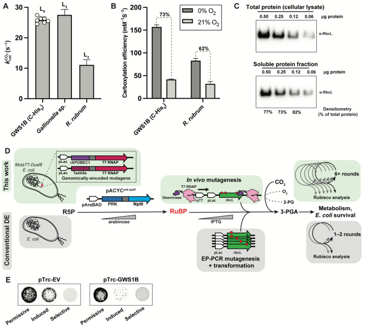
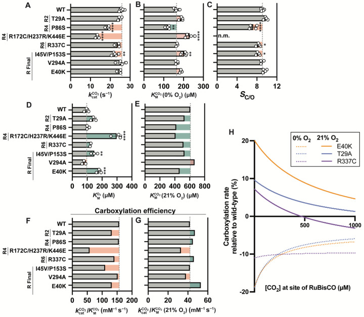
Carbon dioxide (CO2) assimilation by the enzyme Ribulose-1,5-bisphosphate Carboxylase/Oxygenase (Rubisco) underpins biomass accumulation in photosynthetic bacteria and eukaryotes. Despite its pivotal role, Rubisco has a slow carboxylation rate ( k c a t C O 2 ) and is competitively inhibited by oxygen (O2). These traits impose limitations on photosynthetic efficiency, making Rubisco a compelling target for improvement. Interest in Form II Rubisco from Gallionellaceae bacteria, which comprise a dimer or hexamer of large subunits, arises from their nearly 5-fold higher k c a t C O 2 than the average Rubisco enzyme. As well as having a fast k c a t C O 2 (25.8 s -1 at 25 °C), we show that Gallionellaceae Rubisco (GWS1B) is extremely sensitive to O2 inhibition, consistent with its evolution under semi-anaerobic environments. We therefore used a novel in vivo mutagenesis-mediated screening pipeline to evolve GWS1B over six rounds under oxygenic selection, identifying three catalytic point mutants with improved ambient carboxylation efficiency; Thr-29-Ala (T29A), Glu-40-Lys (E40K) and Arg-337-Cys (R337C). Full kinetic characterization showed that each substitution enhanced the CO2 affinity of GWS1B under oxygenic conditions by subduing oxygen affinity, leading to 25% (E40K), 11% (T29A) and 8% (R337C) enhancements in carboxylation efficiency under ambient O2 at 25 °C. By contrast, under the near anaerobic natural environment of Gallionellaceae, the carboxylation efficiency of each mutant was impaired ~16%. These findings demonstrate the efficacy of artificial directed evolution to access novel regions of catalytic space in Rubisco.


Figures

Figure 1.
Figure 1.. Biochemistry of GWS1B Rubisco and potential for in vivo directed evolution in E. coli:
A. Carboxylation rates for C-terminally-tagged GWS1B Rubisco (this work), as well as for Gallionella sp. Rubisco (2) and R. rubrum Rubisco (48). B. Carboxylation efficiencies at 0% O2 and 21% O2 for GWS1B and R. rubrum Rubisco (48). C. Western blot analysis of Rubisco levels detected in total protein lysate (top) compared to the soluble protein fraction (bottom) when GWS1B is expressed from the pTrc-GWS1B plasmid, revealing the very high soluble expression of GWS1B. D. Traditional Rubisco directed evolution workflow versus targeted mutagenesis-mediated in vivo directed evolution workflow. In vivo directed evolution is enabled by in-cell, MutaT7-based mutagenesis rather than by extracellular, error-prone PCR (ep-PCR) mutagenesis followed by transformation into E. coli cells. MutaT7-based mutagenesis, enabled by a deaminase-T7 RNA polymerase (T7 RNAP) fusion binding to and processing from the T7 RNAP promoter site (pT7) on rbcL, generates high levels of downstream mutations in the plasmid DNA and avoids the need for step-wise generation of mutations and transformation, enabling deeper mutagenesis and more rapid progress between rounds of directed evolution. In both workflows, an exogenous prk gene is expressed to create a circuit where E. coli survival is dependent on Rubisco-catalyzed carboxylation and detoxification of RuBP. R5P = Ribulose-5-phosphate; RuBP = Ribulose-1,5-bisphosphate; 2-PG = 2-phosphoglycolate; 3-PGA = 3-phosphoglycerate. E. RDE testing of GWS1B against an empty vector (pTrc-EV) control. Permissive growth media contains no additives, induced growth media contains 0.5 mM IPTG for expression of Rubisco, and selective growth media contains 0.5 mM IPTG, kanamycin at 400 mg/mL, and 0.15% L-arabinose for expression of PRK-NPTII.
Figure 2.
Figure 2.. Identification and biochemical characterization of GWS1B variants:
A. Schematic of MutaT7-RDE workflow. B. Map of the promoter and GWS1B-coding region of the BAC evolution plasmid showing mutational frequency at the DNA base level during sequenced rounds of evolution. Non-synonymous mutations from clonal isolates are indicated with arrows C. Non-synonymous mutations found in clonal isolates of GWS1B in rounds 1, 2, 4, 6, and final rescreening round of evolution. D. Soluble, folded Rubisco content of selected variants in E. coli quantified by [14C]-CABP binding (50). Data are the means and standard deviations of three replicates with significance shown relative to wild-type GWS1B as analyzed by one-way ANOVA. **, p ≤ .005; ***, p ≤ .0005; ****, p ≤ .0001. E. Aggregation temperature (Tagg) of selected variants. Significance testing and labels are consistent with (D). F. SDS-PAGE immunoblots of GWS1B RbcL levels in the soluble and total protein fractions upon expression of GWS1B variants in E. coli.
Figure 3.
Figure 3.. Kinetic characterization of GWS1B variants:
A. Carboxylation rate. B. Affinity for CO2. C. Specificity factor. D. Affinity for O2. Asterisks indicate p-value as calculated by one-way ANOVA followed by a post-hoc Tukey test. *, p ≤ .05; **, p ≤ .005; ***, p ≤ .0005; ****, p ≤ .0001. E. Affinity for CO2in air. F. Carboxylation efficiency at 0% O2. G. Carboxylation efficiency at 21% O2. H. Carboxylation rates of GWS1B E40K, GWS1B T29A, and GWS1B R337C relative to wild-type GWS1B at 0% or 21% O2 as a function of CO2 concentration.
Figure 4.
Figure 4.. Mapping evolved variants onto GWS1B:
A. Substitutions T29A, E40K, and I45V are positioned near the GWS1B active site. PDBID: 5C2G (37). B. E40K, I45V, P86S, R172C, H237R, V294A, and K446E occur at the dimer interface. R337C is surface-exposed. C. P153S occurs at the hexamer interface of GWS1B.

Similar articles

References

    1. Prywes N., Phillips N. R., Tuck O. T., Valentin-Alvarado L. E., Savage D. F., Rubisco function, evolution, and engineering. Annu. Rev. Biochem. 92, 385–410 (2023). - PubMed
    1. Davidi D. et al. , Highly active rubiscos discovered by systematic interrogation of natural sequence diversity. EMBO J. 39, e104081 (2020). - PMC - PubMed
    1. Whitney S. M., Houtz R. L., Alonso H., Advancing our understanding and capacity to engineer Nature’s CO2-sequestering enzyme, Rubisco. Plant Physiol. 155, 27–35 (2011). - PMC - PubMed
    1. Tabita F. R., Satagopan S., Hanson T. E., Kreel N. E., Scott S. S., Distinct form I, II, III, and IV Rubisco proteins from the three kingdoms of life provide clues about Rubisco evolution and structure/function relationships. J. Exp. Bot. 59, 1515–1524 (2008). - PubMed
    1. Schulz L. et al. , Evolution of increased complexity and specificity at the dawn of form I Rubiscos. Science 378, 155–160 (2022). - PubMed

Publication types