Review
. 2025 Jul 8:18:8945-8959.
doi: 10.2147/JIR.S527764. eCollection 2025.

Macrophages as Multifaceted Orchestrators of Tissue Repair: Bridging Inflammation, Regeneration, and Therapeutic Innovation

Affiliations
Review

Macrophages as Multifaceted Orchestrators of Tissue Repair: Bridging Inflammation, Regeneration, and Therapeutic Innovation

Li Wang et al. J Inflamm Res. .

Abstract

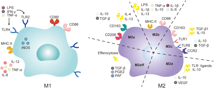
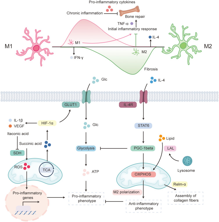
Macrophages play pivotal roles in tissue repair through remarkable functional plasticity, orchestrated by their developmental origins and local microenvironmental cues. Embryonically derived resident macrophages primarily maintain tissue homeostasis, while monocyte-derived macrophages respond predominantly to inflammation and extracellular matrix remodeling. Effective tissue repair requires precise temporal regulation of macrophage polarization, balancing inflammation resolution, angiogenesis, and scar formation. Metabolic reprogramming further enhances macrophage plasticity, enabling adaptation to fluctuating energy demands at injury sites. Emerging evidence also highlights that macrophages integrate biomechanical forces-such as matrix stiffness and shear stress-with biochemical signals to fine-tune their inflammatory and reparative programs. Recognizing this mechanoregulation broadens therapeutic avenues for precisely modulating macrophage behavior in regenerative medicine. Targeting macrophage subsets, polarization states, or metabolic pathways has emerged as a promising therapeutic strategy to optimize healing outcomes. However, the inherent complexity of macrophage heterogeneity presents considerable challenges to therapeutic precision. This review systematically summarizes the multifaceted roles of macrophages in tissue repair, emphasizing how developmental origins dictate functional specificity, dynamic phenotypic transitions, and metabolic adaptability, aiming to advance macrophage-based precision therapeutics for regenerative medicine.

Keywords: macrophage; macrophage polarization; therapeutic strategy; tissue repair.


Conflict of interest statement

The authors report no conflicts of interest in this work.

Figures

Figure 1
Figure 1
Functional Dynamics of Macrophage Polarization in Inflammatory and Tissue Repair Processes. Upon stimulation with LPS, TNF-α, or IFN-γ, M1 macrophages display pro-inflammatory characteristics, marked by elevated expression of TLR-2, TLR-4, CD80, CD86, iNOS, and MHC-II. Activation of NF-κB and STAT1 signaling pathways promotes the release of IL-6, IL-12, and TNF-α, which in turn intensifies inflammation and aids in pathogen elimination during acute wound healing. During tissue repair, macrophages shift toward anti-inflammatory and pro-repair M2 phenotypes. These M2 subtypes are distinguished by specific activation stimuli, surface markers, and functional roles: M2a: Activated by IL-4 or IL-13, these macrophages express CD206 and CD163 while producing IL-10 and TGF-β. M2b: Triggered by immune complexes, LPS, or IL-1β, they retain MHC-II and CD86 expression and secrete TNF-α, IL-1, IL-6, and IL-10. M2c: Stimulated by IL-10 or TGF-β1, they express CD163, CCR2, TLR1, and TLR8 with elevated IL-10 and TGF-β levels. M2d: Activated via TLR ligands or IL-10, they release IL-10 and VEGF. M2eff: Efferocytosis-induced, these macrophages produce TGF-β, PGE2, and PAF.
Figure 2
Figure 2
Metabolic reprogramming of macrophages and their role in tissue repair. M1 macrophages secrete inflammatory cytokines such as TNF-α; the acute inflammatory response they drive promotes bone repair, whereas chronic inflammation impairs regeneration. In contrast, M2 macrophages are detrimental during the early repair phase: their anti-inflammatory mediators (eg, IL-4) suppress necessary early inflammation and thereby inhibit the repair process. Glucose is taken up into macrophages via GLUT1 and metabolized through glycolysis to generate ATP, which drives the pro-inflammatory (M1) phenotype. HIF-1α upregulates GLUT1 expression, further reinforcing this phenotype. Under inflammatory conditions, the TCA‐cycle intermediate succinate accumulates in macrophages; elevated succinate stabilizes HIF-1α and promotes the M1 phenotype. Succinate can also be oxidized by succinate dehydrogenase to produce ROS, which further enhances pro-inflammatory gene expression. Itaconate counteracts this by inhibiting succinate dehydrogenase, thereby blocking succinate’s pro-inflammatory effects. When stimulated with IL-4, macrophages activate the STAT6/PGC-1β pathway to boost mitochondrial oxidative phosphorylation (OXPHOS) and suppress glycolysis; increased OXPHOS favors M2 polarization. Lysosomal acid lipase–mediated lipolysis also supports OXPHOS and M2 differentiation. IL-4–treated macrophages display higher mitochondrial mass, facilitating the release of Relm-α, which signals to fibroblasts to assemble collagen fibers and thereby promotes tissue repair.
Figure 3
Figure 3
Anti-inflammatory and Antifibrotic Functions of Macrophages. High-density lipoprotein (HDL) induces the transcription factor ATF3 in macrophages; ATF3 downregulates Toll-like receptors (TLRs), thereby suppressing TLR-mediated pro-inflammatory cytokine expression. Estrogen-related receptor α (ERRα) binds to the Tnfaip3 promoter, upregulating Tnfaip3 to inhibit TLR-driven inflammation. IL-4 and IL-13 remodel macrophage microRNA profiles—upregulating miR-142-5p and downregulating miR-130a-3p—to target SOCS1 and relieve PPARγ inhibition, which enhances STAT6 phosphorylation and drives expression of pro-fibrotic genes. Macrophages also assemble Connexin 43 (Cx43) gap junctions that allow direct intercellular Ca²+ transfer, activating the Akt pathway in alveolar macrophages (AMs). Via exosomes, macrophages export SOCS1 and SOCS3 to epithelial cells, where they suppress STAT signaling; the combined effect of Akt activation and STAT inhibition alleviates inflammation. Macrophages further promote resolution of inflammation across multiple tissues by regulating regulatory T cells (Tregs) to secrete IL-10 and TGF-β1. Finally, macrophage insulin receptor (IR) and IGF-1 receptor (IGF-1R) signaling play critical roles in controlling interactive inflammatory responses between the epidermis and dermis.

Similar articles

References

    1. Hassanshahi A, Hassanshahi M, Khabbazi S, et al. Adipose-derived stem cells for wound healing. J Cell Physiol. 2019;234(6):7903–7914. doi: 10.1002/jcp.27922 - DOI - PubMed
    1. Gordon S, Martinez FO. Alternative activation of macrophages: mechanism and functions. Immunity. 2010;32(5):593–604. doi: 10.1016/j.immuni.2010.05.007 - DOI - PubMed
    1. Wynn TA. Cellular and molecular mechanisms of fibrosis. J Pathol. 2008;214(2):199–210. doi: 10.1002/path.2277 - DOI - PMC - PubMed
    1. Shapouri-Moghaddam A, Mohammadian S, Vazini H, et al. Macrophage plasticity, polarization, and function in health and disease. J Cell Physiol. 2018;233(9):6425–6440. doi: 10.1002/jcp.26429 - DOI - PubMed
    1. Manole E, Niculite C, Lambrescu IM, et al. Macrophages and stem cells-two to tango for tissue repair? Biomolecules. 2021;11(5):697. doi: 10.3390/biom11050697 - DOI - PMC - PubMed

LinkOut - more resources