Atypical Presentation of Systemic Amyloid Light Chain (AL) Amyloidosis
- PMID: 40656390
- PMCID: PMC12254046
- DOI: 10.7759/cureus.85777
Atypical Presentation of Systemic Amyloid Light Chain (AL) Amyloidosis
Abstract
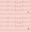
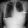
Light chain (AL) amyloidosis is caused by a plasma cell clone that produces a dysfunctional, monoclonal light chain. This light chain misfolds and aggregates, resulting in catastrophic organ damage and eventual death. Due to the multiple organs affected by the disease, symptoms can be vague, such as lethargy, fatigue, weight loss, and peripheral edema. This case presentation follows a 66-year-old female patient who presented to our emergency department complaining of progressive shortness of breath (SOB). The patient denied any other associated symptoms and denied any past significant medical history. The lack of other associated symptoms or past diagnoses was a part of this atypical presentation, as the patient frequently complained of a multitude of symptoms affecting more than one organ system, as the pathogenesis of the disease is systemic in nature. Additionally, individuals with undiagnosed amyloidosis have often been diagnosed with other disease processes based on their symptomatology, due to their sometimes nonspecific nature. Upon further evaluation, bilateral pleural effusions were noted on imaging, which was treated with a pigtail pleural catheter. Atrial fibrillation was noted on ECG and was treated with both rate- and rhythm-controlled amiodarone. AL amyloidosis was suspected on transthoracic cardiac ultrasound, which was described as a "grainy" appearance of the myocardium, along with heart failure and left ventricular hypertrophy, supported by serum protein electrophoresis (SPEP) and urine protein electrophoresis (UPEP) results. This was confirmed by abdominal fat pad and bone marrow biopsies. While the use of cardiac ultrasound in the preliminary workup of amyloidosis is supported by evidence-based practice, there is a need for confirmatory diagnostic testing. This led to the decision to utilize fat pad biopsy as the primary definitive diagnostic tool, as it was readily available within the hospital system and was technically easier than many of the alternatives. However, this is neither supported nor refuted as the gold standard by the available literature. The patient was transferred to a higher level of care at a center specifically focused on the treatment of amyloidosis to receive the current gold standard of treatment, which was unavailable at our facility due to a lack of these specialized medications, as well as a provider comfortable with the use of these treatments.
Keywords: amyloidosis al; cardiac amyloidosis; cardiac amyloidosis with reduced ejection fraction; daratumumab; delayed diagnosis cardiac amyloidosis; light chain amyloidosis.
Copyright © 2025, Faraci et al.
Conflict of interest statement
Human subjects: Consent for treatment and open access publication was obtained or waived by all participants in this study. Conflicts of interest: In compliance with the ICMJE uniform disclosure form, all authors declare the following: Payment/services info: All authors have declared that no financial support was received from any organization for the submitted work. Financial relationships: All authors have declared that they have no financial relationships at present or within the previous three years with any organizations that might have an interest in the submitted work. Other relationships: All authors have declared that there are no other relationships or activities that could appear to have influenced the submitted work.
Figures


Similar articles
-
Signs and symptoms to determine if a patient presenting in primary care or hospital outpatient settings has COVID-19.Cochrane Database Syst Rev. 2022 May 20;5(5):CD013665. doi: 10.1002/14651858.CD013665.pub3. Cochrane Database Syst Rev. 2022. PMID: 35593186 Free PMC article.
-
The Black Book of Psychotropic Dosing and Monitoring.Psychopharmacol Bull. 2024 Jul 8;54(3):8-59. Psychopharmacol Bull. 2024. PMID: 38993656 Free PMC article. Review.
-
Sexual Harassment and Prevention Training.2024 Mar 29. In: StatPearls [Internet]. Treasure Island (FL): StatPearls Publishing; 2025 Jan–. 2024 Mar 29. In: StatPearls [Internet]. Treasure Island (FL): StatPearls Publishing; 2025 Jan–. PMID: 36508513 Free Books & Documents.
-
Systemic Inflammatory Response Syndrome.2025 Jun 20. In: StatPearls [Internet]. Treasure Island (FL): StatPearls Publishing; 2025 Jan–. 2025 Jun 20. In: StatPearls [Internet]. Treasure Island (FL): StatPearls Publishing; 2025 Jan–. PMID: 31613449 Free Books & Documents.
-
Systemic pharmacological treatments for chronic plaque psoriasis: a network meta-analysis.Cochrane Database Syst Rev. 2021 Apr 19;4(4):CD011535. doi: 10.1002/14651858.CD011535.pub4. Cochrane Database Syst Rev. 2021. Update in: Cochrane Database Syst Rev. 2022 May 23;5:CD011535. doi: 10.1002/14651858.CD011535.pub5. PMID: 33871055 Free PMC article. Updated.
References
-
- Cardiac amyloidosis: evolving diagnosis and management: a scientific statement from the American Heart Association. Kittleson MM, Maurer MS, Ambardekar AV, et al. Circulation. 2020;142:0. - PubMed
-
- Serum N-terminal pro-brain natriuretic peptide is a sensitive marker of myocardial dysfunction in AL amyloidosis. Palladini G, Campana C, Klersy C, et al. Circulation. 2003;107:2440–2445. - PubMed
-
- Immunoglobulin light chain amyloidosis: 2020 update on diagnosis, prognosis, and treatment. Gertz MA. Am J Hematol. 2020;95:848–860. - PubMed
Publication types
LinkOut - more resources
Full Text Sources
Miscellaneous
