

Artificial Intelligence in Ultrasound-Based Diagnoses of Gynecological Tumors: A Systematic Review
- PMID: 40656430
- PMCID: PMC12255850
- DOI: 10.7759/cureus.85884
Artificial Intelligence in Ultrasound-Based Diagnoses of Gynecological Tumors: A Systematic Review
Abstract
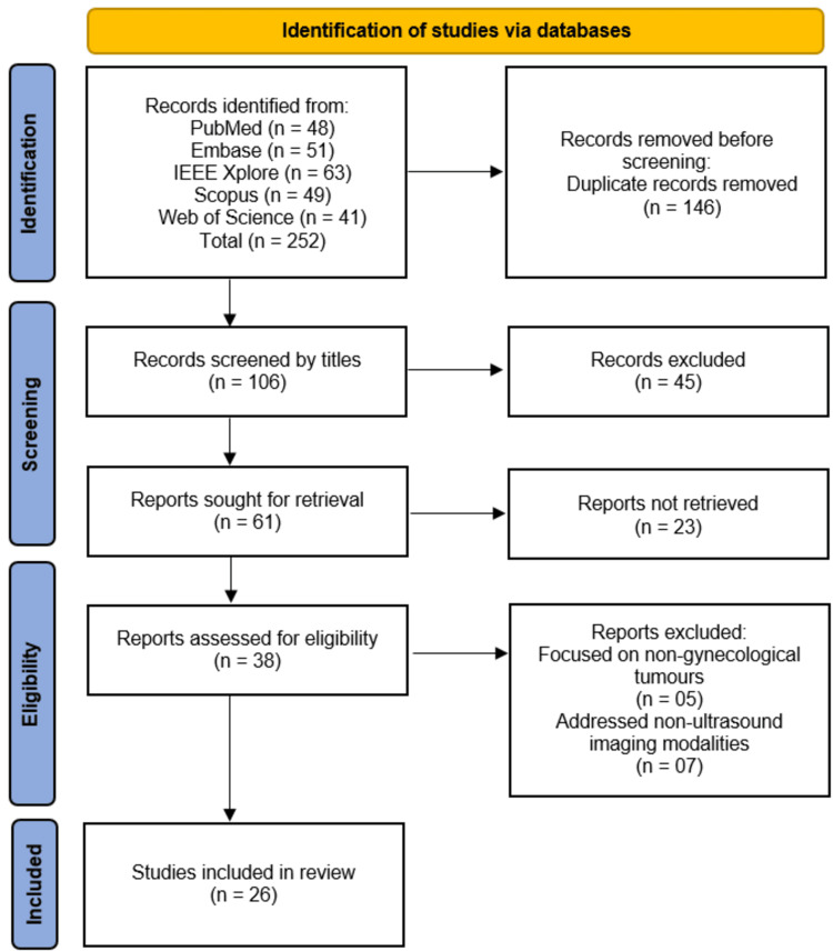
Gynecological tumors, particularly ovarian, endometrial, and uterine masses, pose significant diagnostic challenges due to their heterogeneity and the subjective nature of ultrasound interpretation. Artificial intelligence (AI) has emerged as a promising tool to enhance diagnostic accuracy, yet its clinical adoption remains limited. This systematic review synthesizes evidence on AI applications in ultrasound-based diagnosis of gynecological tumors, evaluating performance metrics, methodological strengths, and limitations to guide future research and clinical implementation. Following Preferred Reporting Items for Systematic Reviews and Meta-Analyses (PRISMA) 2020 guidelines, a comprehensive search was conducted across PubMed, Excerpta Medica Database (Embase), Institute of Electrical and Electronics Engineers Xplore (IEEE Xplore), Scopus, and Web of Science, yielding 252 records. After removing duplicates and screening titles/abstracts, 106 studies were assessed, with 26 meeting inclusion criteria. Eligible studies investigated AI models for gynecological tumor diagnosis using ultrasound. Data were extracted on study design, sample size, AI methodology, performance metrics, and clinical applicability. Risk of bias was assessed using Quality Assessment of Diagnostic Accuracy Studies-2 (QUADAS-2). Narrative synthesis was performed due to methodological heterogeneity. The 26 included studies demonstrated strong diagnostic performance, with AI models achieving accuracies of 75-99.8% and area under the curve (AUCs) up to 0.99 in differentiating benign from malignant tumors. Deep learning architectures (e.g., convolutional neural networks (CNNs), residual neural networks (ResNet)) outperformed traditional machine learning in most studies, particularly when integrating radiomics with clinical variables (e.g., cancer antigen 125 (CA-125)). However, heterogeneity in imaging protocols, sample sizes, and validation methods limited comparability. Only three studies employed prospective designs, and few addressed algorithmic bias or real-world clinical integration. AI shows significant potential to improve ultrasound-based diagnosis of gynecological tumors, offering superior accuracy and reproducibility compared to conventional methods. However, standardized imaging protocols, robust external validation, and prospective trials are needed to translate these tools into clinical practice. Future work should prioritize explainable AI, diverse datasets, and outcome studies to ensure equitable and effective implementation.
Keywords: artificial intelligence; diagnostic accuracy; gynecological tumors; machine learning; systematic review; ultrasound.
Copyright © 2025, Abdalla Mohammed et al.
Conflict of interest statement
Conflicts of interest: In compliance with the ICMJE uniform disclosure form, all authors declare the following: Payment/services info: All authors have declared that no financial support was received from any organization for the submitted work. Financial relationships: All authors have declared that they have no financial relationships at present or within the previous three years with any organizations that might have an interest in the submitted work. Other relationships: All authors have declared that there are no other relationships or activities that could appear to have influenced the submitted work.
Figures
Similar articles
-
Artificial Intelligence Applied to Ultrasound Diagnosis of Pelvic Gynecological Tumors: A Systematic Review and Meta-Analysis.Gynecol Obstet Invest. 2025 May 8:1-22. doi: 10.1159/000545850. Online ahead of print. Gynecol Obstet Invest. 2025. PMID: 40340944 Free PMC article.
-
Clinical Impact of Artificial Intelligence-Based Triage Systems in Emergency Departments: A Systematic Review.Cureus. 2025 Jun 9;17(6):e85667. doi: 10.7759/cureus.85667. eCollection 2025 Jun. Cureus. 2025. PMID: 40642667 Free PMC article. Review.
-
Application of Artificial Intelligence in Pancreatic Cyst Management: A Systematic Review.Cancers (Basel). 2025 Aug 2;17(15):2558. doi: 10.3390/cancers17152558. Cancers (Basel). 2025. PMID: 40805252 Free PMC article. Review.
-
Advancements in Herpes Zoster Diagnosis, Treatment, and Management: Systematic Review of Artificial Intelligence Applications.J Med Internet Res. 2025 Jun 30;27:e71970. doi: 10.2196/71970. J Med Internet Res. 2025. PMID: 40587773 Free PMC article. Review.
-
Artificial intelligence for diagnosing exudative age-related macular degeneration.Cochrane Database Syst Rev. 2024 Oct 17;10(10):CD015522. doi: 10.1002/14651858.CD015522.pub2. Cochrane Database Syst Rev. 2024. PMID: 39417312
References
-
- Deep learning models for early detection of ovarian cancer: A systematic review of ultrasound-based diagnostic tools. Olosunde AO, Onuiri EE. Asian Journal of Engineering and Applied Technology. 2024;13:15–21.
-
- Artificial intelligence in the diagnosis and management of gynecologic cancer. Paiboonborirak C, Abu-Rustum NR, Wilailak S. Int J Gynaecol Obstet. 2025 - PubMed
-
- Logistic models and artificial intelligence in the sonographic assessment of adnexal masses - a systematic review of the literature. Grigore M, Popovici RM, Gafitanu D, Himiniuc L, Murarasu M, Micu R. Med Ultrason. 2020;22:469–475. - PubMed
Publication types
LinkOut - more resources
Full Text Sources
Research Materials
Miscellaneous