Craniocervical intradural pseudotumor causing bulbomedullary compression
- PMID: 40656481
- PMCID: PMC12255219
- DOI: 10.25259/SNI_403_2025
Craniocervical intradural pseudotumor causing bulbomedullary compression
Abstract
Background: Pseudotumors are rare lesions that may cause compression of adjacent neural structures. Treatment options range from conservative management to surgical intervention.
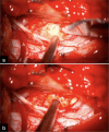
Case description: A 59-year-old female presented with a 3-month history of headaches, difficulty speaking, swallowing, gait disturbance, and progressive left-sided weakness. Her examination confirmed left-sided tetraparesis. The cervical magnetic resonance showed a right-sided mass compressing the bulbomedullary junction. Through a modified right-sided far lateral craniotomy, an intradural "pseudotumor" was removed. Postoperatively, the patient's symptoms gradually improved. Histopathological analysis revealed an acellular fibrocartilaginous mass consistent with the diagnosis of pseudotumor.
Conclusion: Pseudotumors at the craniocervical junction may cause progressive tetraparesis readily resolved following gross total surgical excision.
Keywords: Bulbomedullary compression; Craniocervical pseudotumor; Far lateral craniotomy; Myelopathy.
Copyright: © 2025 Surgical Neurology International.
Conflict of interest statement
There are no conflicts of interest.
Figures


Similar articles
-
No Difference in Revision Rates and High Survival Rates in Large-head Metal-on-metal THA Versus Metal-on-polyethylene THA: Long-term Results of a Randomized Controlled Trial.Clin Orthop Relat Res. 2024 Jul 1;482(7):1173-1182. doi: 10.1097/CORR.0000000000002924. Epub 2023 Dec 12. Clin Orthop Relat Res. 2024. PMID: 38084856 Free PMC article. Clinical Trial.
-
Change in the Location of a Pseudotumor Around the C2 Odontoid Process from Posterior to Anterior to the Odontoid Process in the Natural Course: A Case with "Antero-Odontoid Pseudotumor" or "Peri-Odontoid Pseudotumor".J Clin Med. 2025 Jun 12;14(12):4182. doi: 10.3390/jcm14124182. J Clin Med. 2025. PMID: 40565927 Free PMC article.
-
A rare case of intradural intramedullary cervical spinal neurenteric cyst in an adult: illustrative case.J Neurosurg Case Lessons. 2025 Jun 30;9(26):CASE25253. doi: 10.3171/CASE25253. Print 2025 Jun 30. J Neurosurg Case Lessons. 2025. PMID: 40587885 Free PMC article.
-
Frequency, timing, and predictors of neurological dysfunction in the nonmyelopathic patient with cervical spinal cord compression, canal stenosis, and/or ossification of the posterior longitudinal ligament.Spine (Phila Pa 1976). 2013 Oct 15;38(22 Suppl 1):S37-54. doi: 10.1097/BRS.0b013e3182a7f2e7. Spine (Phila Pa 1976). 2013. PMID: 23963005
-
Anterior cervical intradural arachnoid cyst, a rare cause of spinal cord compression: a case report with video systematic literature review.Eur Spine J. 2016 May;25 Suppl 1:19-26. doi: 10.1007/s00586-015-4026-7. Epub 2015 May 21. Eur Spine J. 2016. PMID: 25995113
References
-
- Adada B, Silva MA, Darwish H, Dakwar E. Far-lateral trans-atlas extradural resection of retro-odontoid synovial cyst: Surgical technique and review of literature. Interdiscip Neurosurg. 2019;17:28–35.
-
- Batista AV, Aguiar GB, Daniel JW, Veiga JC. Retro-odontoid pseudotumor: A poorly recognized alteration of the craniocervical junction. Rev Assoc Méd Bras (1992) 2020;66:507–11. - PubMed
-
- Frempong-Boadu AK, Faunce WA, Fessler RG. Endoscopically assisted transoral-transpharyngeal approach to the craniovertebral junction. Neurosurgery. 2002;51:S60–6. - PubMed
Publication types
LinkOut - more resources
Full Text Sources
