. 2025 Mar 15;14(6):103495.
doi: 10.1016/j.eats.2025.103495. eCollection 2025 Jun.

Circumferential Meniscal Reconstruction Using the Semitendinosus Tendon for a Medial Meniscal Posterior Root Tear

Affiliations

Circumferential Meniscal Reconstruction Using the Semitendinosus Tendon for a Medial Meniscal Posterior Root Tear

Takuya Ohno et al. Arthrosc Tech. .

Abstract

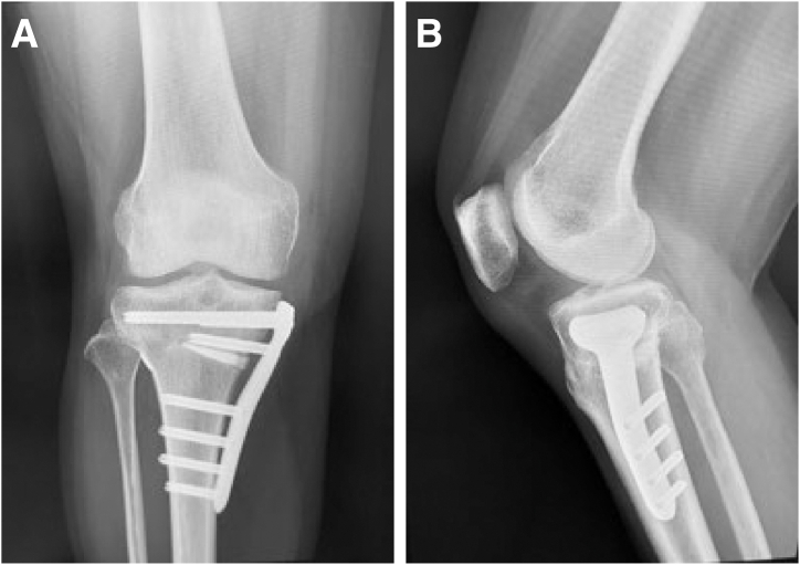
This article describes a surgical technique that addresses the limitations of existing approaches in managing meniscal extrusion after medial meniscal posterior root tear repair. Although traditional methods such as pullout repair and suture anchor repair are highly effective, they often struggle to adequately prevent meniscal extrusion, leading to suboptimal meniscal function restoration. Our method, which uses the semitendinosus tendon for circumferential joint capsule reinforcement, significantly reduces meniscal extrusion and enhances knee stability in patients with medial meniscal posterior root tears. Preliminary results suggest that this technique is superior to conventional methods in terms of preventing meniscal extrusion and restoring knee function.


Conflict of interest statement

All authors (T.O., K.N., H.I., H.O., S.F., M.T., Y.K.) declare that they have no known competing financial interests or personal relationships that could have appeared to influence the work reported in this paper.

Figures

Fig 1
Fig 1
Medial meniscal posterior root tear (MMPRT) in right knee with patient in supine position. (A) An arthroscope is inserted through the anterolateral portal, and the MMPRT is identified and verified. (B) A surgical instrument such as a pair of scissors is used to create a hole in the posterior joint capsule approximately 1.5 cm from the attachment region of the medial meniscal posterior root. An Ethibond is passed through and brought outside the joint capsule. (MFC, medial femoral condyle; MTP, medial tibial plateau.)
Fig 2
Fig 2
The 26-cm semitendinosus tendon is harvested and folded in half. The folded tendon is secured with Ethibond. To construct an extensive graft width, baseball-stitch sutures using Ti-Cron, coated with braided polyester, are placed at each end of the tendon’s double fold and connected side to side with Vicryl suture.
Fig 3
Fig 3
Right knee in supine position from anterolateral portal view. The tibial bone tunnel is formed with a 6.0-mm retrograde drill (AI Drill). The entrance of the bone tunnel should be addressed on medial meniscal posterior root attachment. (MFC, medial femoral condyle; MTP, medial tibial plateau.)
Fig 4
Fig 4
Right knee in supine position from anterolateral portal view. (A) The graft’s Ethibond is drawn out through the anteromedial portal and tied with the Ethibond from the tibial tunnel. By pulling the Ethibond exiting from the tibial tunnel, the graft is relayed into the tunnel using the shuttle-relay method. (B) Fixation is performed with an assist suture using Ultrabraid to facilitate adhesion with the graft and meniscus. (MFC, medial femoral condyle; MTP, medial tibial plateau.)
Fig 5
Fig 5
Right knee in supine position. (A) Medial outside view showing process of pulling Ethibond departing from tibial bone tunnel while making 1-cm incision in tibialis anterior muscle, securing pullout button (AI-Medic), routing graft circumferentially along joint capsule, and applying moderate tension. (B) Visual representation of reconstruction technique.
Fig 6
Fig 6
Postoperative radiographic images of right knee. (A) Frontal view. (B) Lateral view. The pullout button (AI-Medic) is secured on the lateral side of the tibia.
Fig 7
Fig 7
Preoperative (A) and postoperative (B) magnetic resonance images of right knee. Postoperatively, as indicated by the white arrows, the extrusion of the medial meniscus is corrected, and the graft created on the lateral side of the joint capsule is visible.

Similar articles

References

    1. Guermazi A., Hayashi D., Jarraya M., et al. Medial posterior meniscal root tears are associated with development or worsening of medial tibiofemoral cartilage damage: The Multicenter Osteoarthritis study. Radiology. 2013;268:814–821. - PMC - PubMed
    1. Mohamadi A., Momenzadeh K., Masoudi A., et al. Evolution of knowledge on meniscal biomechanics: A 40 year perspective. BMC Musculoskelet Disord. 2021;22:625. - PMC - PubMed
    1. Fox A.J.S., Bedi A., Rodeo S.A. The basic science of human knee menisci: Structure, composition, and function. Sports Health. 2012;4:340–351. - PMC - PubMed
    1. LaPrade C.M., James E.W., Cram T.R., Feagin J.A., Engebretsen L., LaPrade R.F. Meniscal root tears: A classification system based on tear morphology. Am J Sports Med. 2015;43:363–369. - PubMed
    1. Allaire R., Muriuki M., Gilbertson L., Harner C.D. Biomechanical consequences of a tear of the posterior root of the medial meniscus. Similar to total meniscectomy. J Bone Joint Surg Am. 2008;90:1922–1931. - PubMed

LinkOut - more resources