

Prostate cancer classification using 3D deep learning and ultrasound video clips: a multicenter study
- PMID: 40657247
- PMCID: PMC12245699
- DOI: 10.3389/fonc.2025.1582035
Prostate cancer classification using 3D deep learning and ultrasound video clips: a multicenter study
Abstract
Objective: This study aimed to evaluate the effectiveness of deep-learning models using transrectal ultrasound (TRUS) video clips in predicting prostate cancer.
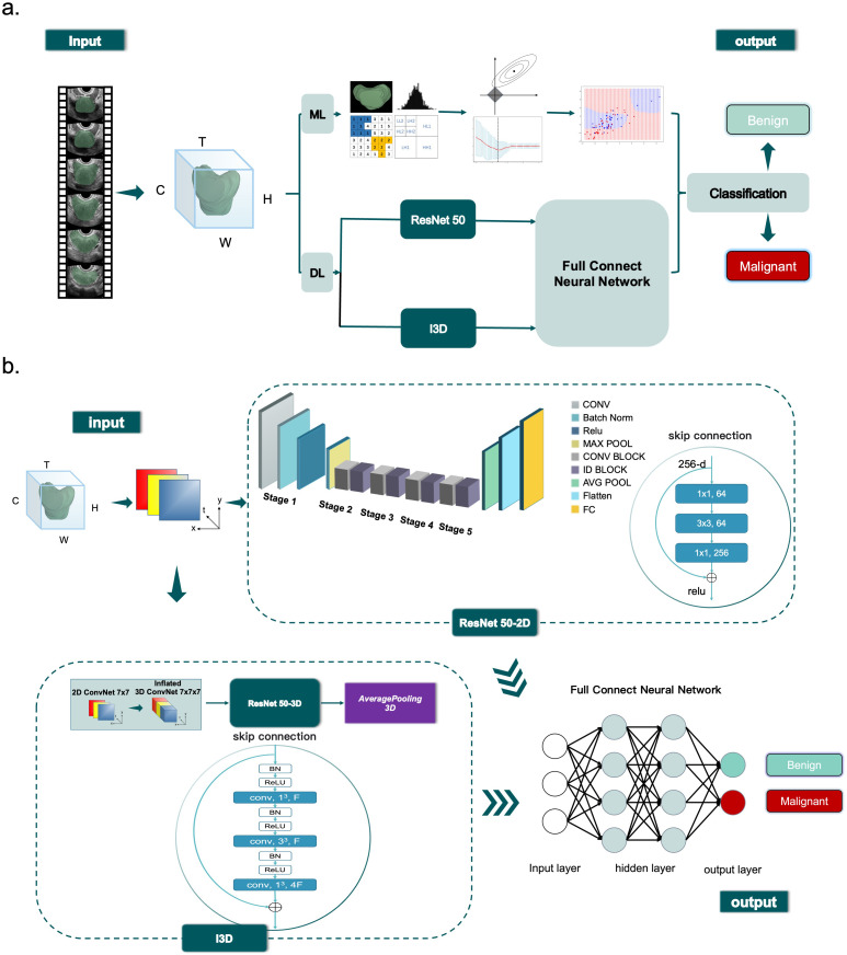
Methods: We manually segmented TRUS video clips from consecutive men who underwent examination with EsaoteMyLab™ Class C ultrasonic diagnostic machines between January 2021 and October 2022. The deep learning-inflated 3D ConvNet (I3D) model was internally validated using split-sample validation on the development set through cross-validation. The final performance was evaluated on two external test sets using geographic validation. We compared the results obtained from a ResNet 50 model, four ML models, and the diagnosis provided by five senior sonologists.
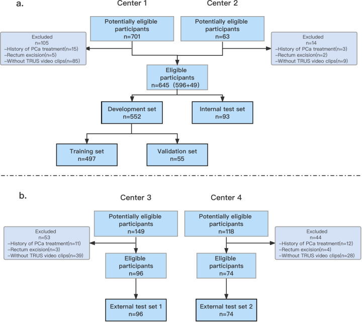
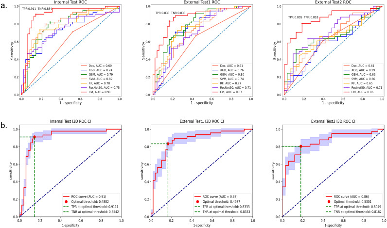
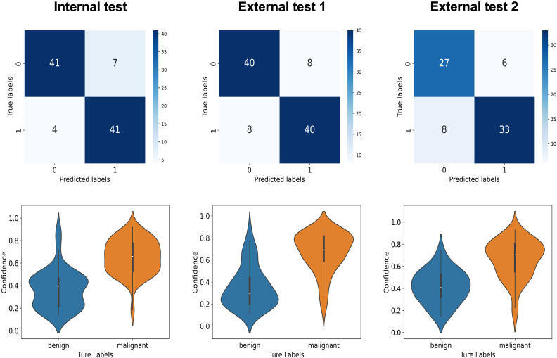
Results: A total of 815 men (median age: 71 years; IQR: 67-77 years) were included. The development set comprised 552 men (median age: 71 years; IQR: 67-77 years), the internal test set included 93 men (median age: 71 years; IQR: 67-77 years), external test set 1 consisted of 96 men (median age: 70 years; IQR: 65-77 years), and external test set 2 had 74 men (median age: 72 years; IQR: 68-78 years). The I3D model achieved diagnostic classification AUCs greater than 0.86 in the internal test set as well as in the independent external test sets 1 and 2. Moreover, it demonstrated greater consistency in sensitivity, specificity, and accuracy compared to pathological diagnosis (kappa > 0.62, p < 0.05). It exhibited a statistically significant superior ability to classify and predict prostate cancer when compared to other AI models, and the diagnoses provided by sonologists (p<0.05).
Conclusion: The I3D model, utilizing TRUS prostate video clips, proved to be valuable for classifying and predicting prostate cancer.
Keywords: I3D model; deep learning; multicenter study; prostate cancer; ultrasound.
Copyright © 2025 Lou, Chen, Wu, Liu, Zhou, Zhang, Tu, Hu, Lv, Yang, Qi, Sun, Du, Liu, Zhou, Liu, Chen, Wang, Yao and Wang.
Conflict of interest statement
The authors declare that the research was conducted in the absence of any commercial or financial relationships that could be construed as a potential conflict of interest.
Figures
References
-
- Mottet N, Van den Bergh RCN, Briers E, Van den Broeck T, Cumberbatch MG, De Santis M, et al. EAU-EANM-ESTRO-ESUR-SIOG guidelines on prostate cancer-2020 update. part 1: Screening, diagnosis, and local treatment with curative intent. Eur Urol. (2021) 79:243–62. doi: 10.1016/j.eururo.2020.09.042 - DOI - PubMed
-
- Olaf R, Philipp F, Thomas B. U-net: convolutional networks for biomedical image segmentation. MICCAI. (2015), 397–405. doi: 10.48550/arXiv.1505.04597 - DOI
LinkOut - more resources
Full Text Sources