. 2025 Jun 23;16(9):2890-2902.
doi: 10.7150/jca.111555. eCollection 2025.

Radioresistant triple-negative breast cancer cells release β-catenin containing extracellular vesicles to promote cancer stem cell activity of bystanders

Affiliations

Radioresistant triple-negative breast cancer cells release β-catenin containing extracellular vesicles to promote cancer stem cell activity of bystanders

Yueh-Chun Lee et al. J Cancer. .

Abstract

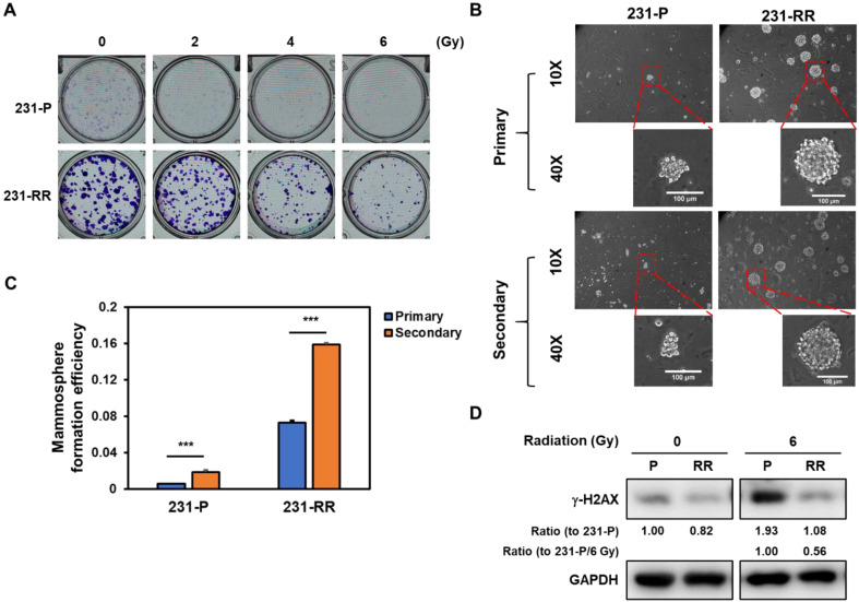
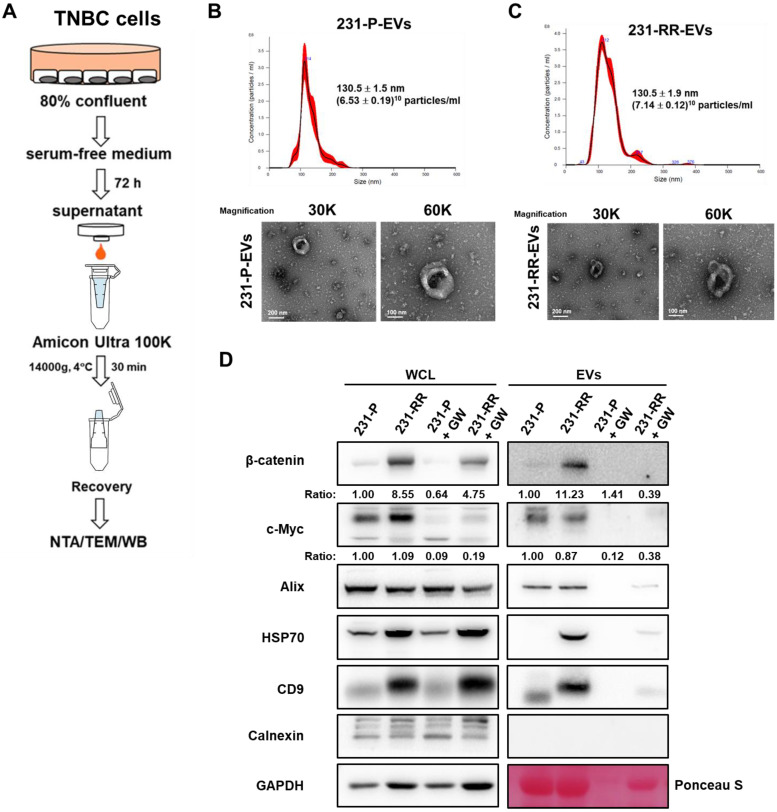
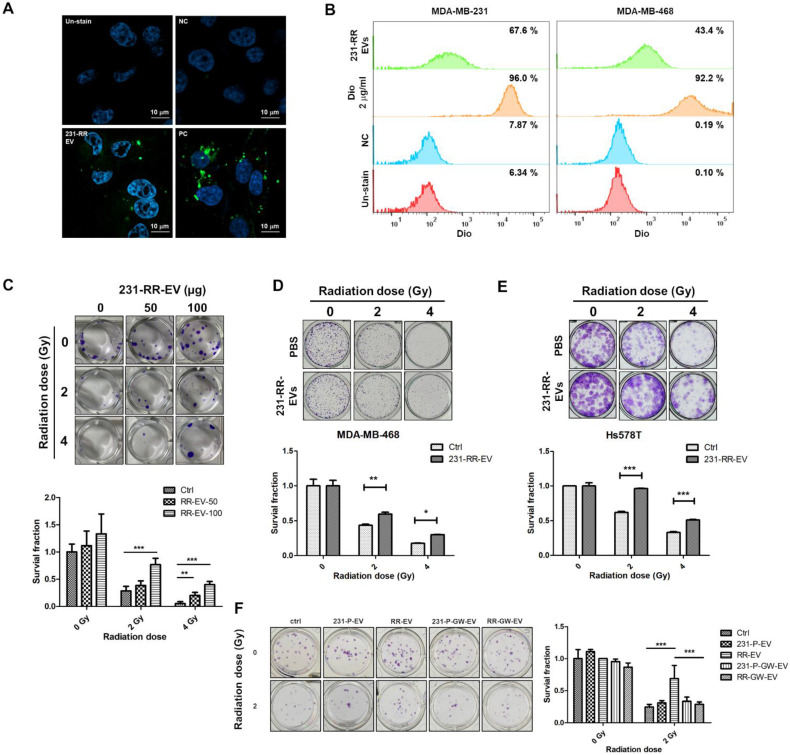
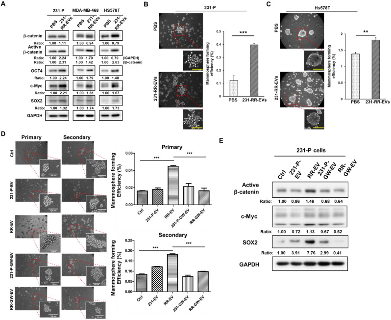
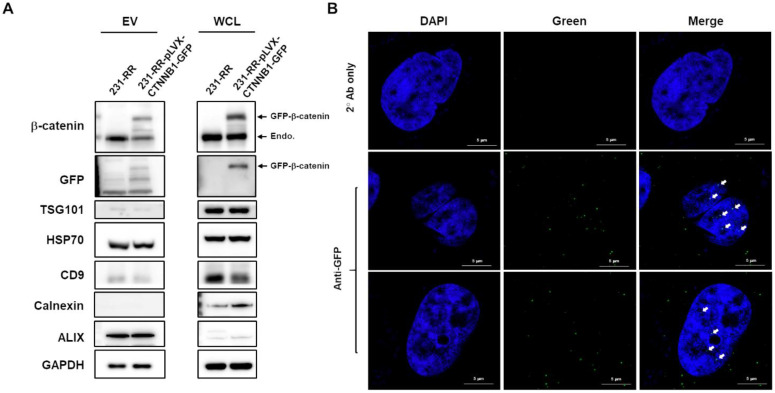
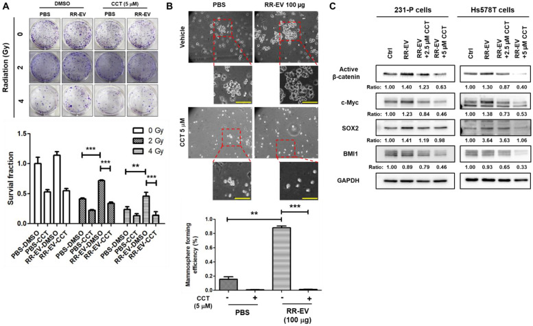
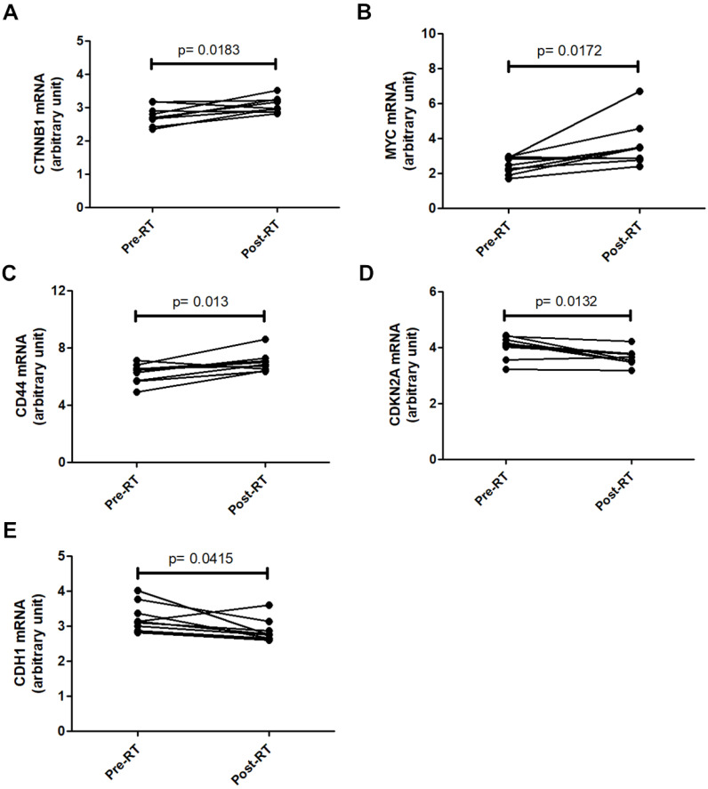
Background: Triple-negative breast cancer (TNBC) frequently develops radioresistance, yet the mechanisms remain incompletely elucidated. This study is the first to investigate how β-catenin, transported by extracellular vesicles (EVs) from radioresistant TNBC cells, promotes radioresistance and enhances cancer stem cell (CSC) activity in recipient TNBC cells, offering a novel mechanism distinct from prior EV-related findings in other cancers. Methods and Results: A radioresistant cell line (231-RR) was developed from MDA-MB-231 cells, and EVs were isolated for characterization. EVs from 231-RR cells decreased radiosensitivity in parental MDA-MB-231 and two other TNBC cell lines (MDA-MB-468 and Hs578T), as shown by clonogenic assay. These EVs also enhanced CSC activity in MDA-MB-231 and Hs578T cells, demonstrated through primary and secondary mammosphere formation. The effects were nullified when using EVs from 231-RR cells treated with the EV secretion inhibitor GW4869. 231-RR-derived EVs showed elevated β-catenin levels and increased active β-catenin and stemness proteins (c-Myc, OCT4, SOX2) in recipient TNBC cells. The β-catenin inhibitor CCT-031374 prevented EV-mediated enhancement of radioresistance and CSC activity. Public data analysis from breast cancer patients revealed post-radiotherapy upregulation of the β-catenin pathway, with elevated CTNNB1, MYC, and CD44 expression, alongside reduced CDKN2A and CDH1 levels, supporting clinical relevance. Conclusions: This study uniquely demonstrates that EVs from radioresistant TNBC cells transfer β-catenin to confer radioresistance and enhance CSC activity in recipient cells, a mechanism not previously reported in TNBC. These findings suggest the potential of EV-β-catenin derived as a novel biomarker for predicting radiotherapy outcomes and recurrence risk in TNBC patients, pending development of sensitive detection methods.

Keywords: cancer stem cells; extracellular vesicles; radioresistance; triple negative breast cancer; β-catenin.


Conflict of interest statement

Competing Interests: The authors have declared that no competing interest exists.

Figures

Figure 1
Figure 1
Radioresistant MDA-MB-231 cells exhibit enhanced radioresistance, elevated CSC activity, and reduced irradiation-induced DNA damage. (A) Clonogenic survival assay comparing parental MDA-MB-231 (231-P) and radioresistant MDA-MB-231 cells (231-RR) following exposure to increasing doses of ionizing radiation (0-6 Gy). Representative images of crystal violet-stained colonies are shown for each condition. (B, C) Representative images of primary and secondary tumorspheres are shown in (B). Each panel includes both low magnification (10× objective) and high magnification (40× objective) views. Enlarged tumorspheres correspond to regions marked with red dashed boxes in the low-magnification images. Scale bar = 100 µm in high-magnification images. Quantification of primary and secondary tumorsphere formation in 231-P and 231-RR cells are shown in (C). Data are presented as mean ± SD. ***, P < 0.001 compared to 231-P. (D) Western blot analysis of γ-H2AX expression in 231-P and 231-RR cells as a marker of DNA double-strand breaks following radiation exposure.
Figure 2
Figure 2
EVs released from radioresistant MDA-MB-231 cells contain high levels of β-catenin protein. (A) Schematic flowchart illustrating the collection and isolation EVs from TNBC cells. NTA, nanoparticle tracking analysis; TEM, transmission electron microscope; WB, western blot. (B, C) Characterization of EVs from 231-P (B) and 231-RR (C) by NTA (upper panels) and TEM (lower panels). Mean particle size and concentration (± SD) are indicated. Representative TEM images of EVs are shown at 30,000× and 60,000× magnification. Scale bars: 200 nm or 100 nm as indicated. (D)Western blot analysis of EV markers, including Alix, HSP70, and CD9, confirming positive identification of EVs. Calnexin, a negative marker for EVs, was included to verify the absence of non-EV contaminants. GAPDH was used as a control for whole cell lysates (WCL), and Ponceau S staining of the EV blot membrane served as a loading control. EVs were also examined from cells treated with 1 µM GW4869 (GW), an inhibitor of EV formation.
Figure 3
Figure 3
EVs from radioresistant MDA-MB-231 cells induce a radioresistant phenotype. (A, B) Extracellular vesicles from radioresistant MDA-MB-231 cells (231-RR-EVs) were labeled with the lipophilic green fluorescent dye DiO. Uptake of 20 µg DiO-labeled 231-RR-EVs by parental MDA-MB-231 cells was examined using confocal microscopy (A) and flow cytometry (B). MDA-MB-468 cell uptake was also analyzed by flow cytometry (B). Cells directly labeled with DiO served as a positive control (PC). PBS with the addition of 2 ug/ml Dio dye at a concentration of 2 ug/ml was also filtered through a 100KDa Amicon Ultra centrifugal filter to dead volume and collected as a negative control (NC). (C) The effect of 231-RR-EVs on the radiosensitivity of parental MDA-MB-231 cells was assessed using a clonogenic assay. MDA-MB-231 cells were treated with 50 µg or 100 µg of 231-RR-EVs for 24 hours prior to exposure to the indicated doses of radiation. (D, E) MDA-MB-468 (D) or Hs578T (E) cells were treated with 231-RR-EVs at a concentration of 100 µg followed by irradiation at the indicated doses. The radiosensitivity was determined by clonogenic assay. (F) MDA-MB-231 cells were treated with EVs from parental cells (231-P-EV), radioresistant cells (RR-EV), or from GW4869-treated cells (231-P-GW-EV, RR-GW-EV) at a concentration of 100 µg for 24 hours, followed by irradiation at 2 Gy. Colonies were visualized by crystal violet staining and counted 14 days post-seeding. Data are presented as relative colony numbers compared to the untreated control (Ctrl) without irradiation. *, P < 0.05; **, P < 0.01; ***, P < 0.001.
Figure 4
Figure 4
EVs from radioresistant MDA-MB-231 cells enhance the expression of cancer stemness proteins and CSC activity. (A) Parental MDA-MB-231 (231-P), MDA-MB-468, or Hs578T cells were treated with 100 µg of EVs derived from radioresistant MDA-MB-231 cells (231-RR-EVs) for 48 hours. The expression of cancer stemness-related proteins, including β-catenin, active β-catenin, OCT4, c-Myc, and SOX2, was analyzed by western blot. (B, C) Parental MDA-MB-231 (B) or Hs578T (C) cells were treated with 100 µg of EVs isolated from 231-P cells (231-P-EVs) for 24 hours. Representative images of mammospheres are shown (left), along with quantification of mammosphere formation efficiency (right). Each image panel includes both low magnification (20× objective) and high magnification (40× objective) images. Enlarged views correspond to areas marked with red dashed boxes in the low-magnification images. Scale bar = 100 µm in high-magnification images. **, P< 0.01; ***, P< 0.001. (D) Parental 231-P cells were treated with EVs from 231-P, 231-RR, or GW4869-treated cells (231-P-GW-EVs or RR-GW-EVs), followed by primary mammosphere formation. After 7 days, primary mammospheres were collected using 100 µm strainers, dissociated with Accutase, and single-cell suspensions were used for secondary mammosphere formation assays. Representative images include low magnification (10× objective) and high magnification (40× objective) views, with enlarged images highlighting areas marked with red dashed boxes. Scale bar = 100 µm in high-magnification images. Quantification of both primary and secondary mammosphere formation efficiency is shown. ***, P < 0.001. (E) The protein expression levels of active β-catenin, c-Myc, and SOX2 were assessed by western blot in 231-P cells treated with 100 µg EVs of 231-P, 231-RR, or GW4869 treated cells (231-P-GW-EVs or RR-GW-EVs) for 48 hours. Ratios indicated the relative expression compared to untreated control cells (ctrl).
Figure 5
Figure 5
Nuclear delivery of β-catenin by EVs released from radioresistant MDA-MB-231 cells. (A) MDA-MB-231 cells (231-RR) were transfected with a plasmid encoding GFP-tagged β-catenin (pLVX-CTNNB1-GFP) for 24 hours, followed by puromycin selection. Whole cell lysates (WCL) and EVs released from 231-RR cells, with or without GFP-tagged β-catenin expression, were collected. Western blot analysis was performed to detect the indicated proteins. (B) Parental MDA-MB-231 cells were treated with 100 µg of EVs from 231-RR cells expressing GFP-tagged β-catenin for 2 hours. GFP expression was detected by immunocytochemical staining using an anti-GFP antibody, followed by confocal microscopy with a magnification of 630X. Arrows indicate positive GFP signals within the cell nucleus. FITC-labeled anti-mouse IgG was used as a control. Scale bars, 5 μm.
Figure 6
Figure 6
Inhibition of β-catenin activity abolishes the effects of EVs from radioresistant cells. Parental MDA-MB-231 cells were treated with 100 µg of EVs from radioresistant MDA-MB-231 cells (RR-EV). (A) After 24 hours of treatment, cells were irradiated with 2 or 4 Gy, and colony-forming ability was assessed by clonogenic assay with or without the β-catenin inhibitor CCT-031374 (CCT) at a concentration of 5 µM. **, P< 0.01; ***P < 0.001. (B) After 24 hours of treatment, CSC activity was measured by mammosphere formation assay with the CCT-031374 treatment (CCT, 5 µM). 0.1% DMSO was used as the vehicle control. Each image panel includes both low magnification (20× objective) and high magnification (40× objective) images. Enlarged views correspond to areas marked with red dashed boxes in the low-magnification images. Scale bar = 100 µm in high-magnification images. **, P< 0.01; ***P < 0.001. Scale bars, 100 μm. (C) After 48 hours of EV treatment in MDA-MB-231 (231-P) or Hs578T cells, with or without CCT-031374 at concentrations of 2.5 µM or 5 µM, total cellular proteins were extracted, and the expression levels of active β-catenin, BMI1, c-Myc, and SOX2 were analyzed by western blotting. GAPDH was used as a house keeping gene. Inserted numbers represent as relative levels compared to non-treated cells.
Figure 7
Figure 7
Changes in β-catenin-related gene expression in breast cancer tissues after radio-therapy. Gene expression data from the GSE59733 dataset were obtained from the GEO database. Expression levels of CTNNB1 (A), MYC (B), and CD44 (C), which are upregulated by β-catenin activation, were compared between breast cancer tissue samples collected before (Pre-RT) and after (Post-RT) radiotherapy. In addition, the expression levels of CDKN2A (D) and CDH1 (E), which are downregulated by β-catenin activation, were similarly compared. Statistically significant differences in expression levels are indicated with p-values.

Similar articles

References

    1. Onitilo AA, Engel JM, Greenlee RT, Mukesh BN. Breast cancer subtypes based on ER/PR and Her2 expression: comparison of clinicopathologic features and survival. Clin Med Res. 2009;7:4–13. - PMC - PubMed
    1. Almansour NM. Triple-Negative Breast Cancer: A Brief Review About Epidemiology, Risk Factors, Signaling Pathways, Treatment and Role of Artificial Intelligence. Front Mol Biosci. 2022;9:836417. - PMC - PubMed
    1. Bianchini G, Balko JM, Mayer IA, Sanders ME, Gianni L. Triple-negative breast cancer: challenges and opportunities of a heterogeneous disease. Nat Rev Clin Oncol. 2016;13:674–90. - PMC - PubMed
    1. Kumar P, Aggarwal R. An overview of triple-negative breast cancer. Arch Gynecol Obstet. 2016;293:247–69. - PubMed
    1. Haffty BG, Yang Q, Reiss M, Kearney T, Higgins SA, Weidhaas J. et al. Locoregional relapse and distant metastasis in conservatively managed triple negative early-stage breast cancer. J Clin Oncol. 2006;24:5652–7. - PubMed

LinkOut - more resources