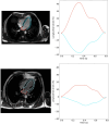
Deep learning-based segmentation and strain analysis of left heart chambers from long-axis CMR images
- PMID: 40657429
- PMCID: PMC12256142
- DOI: 10.1093/ehjimp/qyaf070
Deep learning-based segmentation and strain analysis of left heart chambers from long-axis CMR images
Abstract
Aims: Feature tracking (FT) is increasingly used on dynamic cardiac magnetic resonance (CMR) images for myocardial strain evaluation but often requires manual initialization, which is tedious and source of variability, especially on the challenging long-axis (LAX) images. Accordingly, we designed a pipeline combining deep learning (DL) with FT for left ventricular (LV) and left atrial (LA) longitudinal myocardial strain estimation.
Methods and results: We studied a multivendor database of 684 individuals divided into: training = 845, tuning = 281, and testing = 116 LAX-CMR cine 2- and/or 4-chamber views. Images were centre cropped. Then, a 2D- and 3D-ResUnet, which considers time as the third dimension, were designed for LV/LA segmentation and used to (i) estimate LV and LA strains (Full 2D-/3D-DL) and (ii) initialize an FT algorithm and further derive LV and LA strains (FT-initialized by 2D-/3D-DL). Left ventricular and LA contours and strain peaks were compared against reference standard (RS) measures performed by an expert using a semiautomated software. Intraclass-correlation-coefficient (ICC) was used to study reproducibility. 3D-DL outperformed 2D-DL segmentation (Dice-scores: 0.94 ± 0.02 vs. 0.90 ± 0.09, P = 0.002) and was stable across vendors, field strengths and imaging views. The added value of combining DL with FT was revealed by higher correlations and lower Bland-Altman biases against RS for FT initialized by 3D-DL strains (r ≥ 0.91, |mean-bias|≤0.65%) than for full 3D-DL strains (r ≤ 0.80, |mean-bias|<3.07%). Semiautomated human vs. FT initialized by 3D-DL (ICC ≥ 0.76) and inter-human strain reproducibility was equivalent.
Conclusion: Generalizable DL-based LV and LA segmentation on LAX-CMR images was proposed. Its combination with FT resulted in fully automated and reliable LV and LA strain measures, reaching human reproducibility.
Keywords: CMR; deep learning; feature tracking; left atrium; longitudinal strain.
© The Author(s) 2025. Published by Oxford University Press on behalf of the European Society of Cardiology.
Conflict of interest statement
Conflict of interest: None declared.
Figures


References
LinkOut - more resources
Full Text Sources
Miscellaneous
