. 2025 Jul 1;66(9):37.
doi: 10.1167/iovs.66.9.37.

Longitudinal Study of Changes in Retinal Curvature and its Relationship With Myopia Shift in Chinese Children

Affiliations

Longitudinal Study of Changes in Retinal Curvature and its Relationship With Myopia Shift in Chinese Children

Yue Wu et al. Invest Ophthalmol Vis Sci. .

Abstract

Purpose: The purpose of this study was to investigate longitudinal changes in retinal curvature (RC) among Chinese children and its relationship with myopia progression.

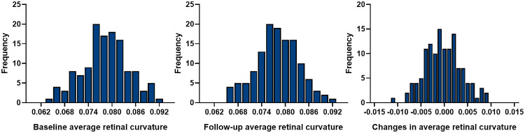
Methods: This 12-month longitudinal study included children aged 6 to 16 years, divided into groups based on changes in spherical equivalent refraction (SER) over 1 year: the myopic shift group and the non-myopic shift group. Comprehensive examinations including swept-source optical coherence tomography were performed in each visit. RC was assessed using a customized 3D reconstruction algorithm. The central, temporal, nasal, superior, and inferior quadrates were further divided into nine regions and calculated the average curvature for each. The retinal surface asymmetry index (R-SAI) was calculated to describe regional differences in RC. Statistical analyses were performed to explore correlations between RC changes and myopia progression.

Results: Significant increases in central RC were observed in the myopic shift group, particularly in the 0 to 6 mm-diameter central retinal region. Regional analysis showed that significant changes in curvature were observed in the myopic shift group particularly in the 0 to 3 mm central circle (C0), temporal quadrate of 3 to 6 mm (T1), and 6 to 9 mm ring (T2) and inferior quadrate of 3 to 6 mm ring (I1), with the most predominant increase in the T1 region. Negative correlation was found between baseline nasal-temporal R-SAI and myopic shift after controlling confounders. Age, axial length (AL) elongation, and smaller baseline curvature were associated with larger RC changes in the central macular region.

Conclusions: RC changes are closely linked to myopia shift. Longitudinal monitoring of RC can serve as a quantitative measure for assessing morphological changes associated with myopia shift in children.


Conflict of interest statement

Disclosure: Y. Wu, None; X. Liu, None; W. Qian, None; Y. Yu, None; S. Wu, None; B. Ni, None; B. Ke, None

Figures

Figure 1.
Figure 1.
An illustration of the difference between uncorrected and corrected OCT. (A) OCT image is processed and presented in a rectangular format. (B) OCT demonstrated a fan-shape scanning beam. All light scans share a common pivot at the nodal point in an optical system. According to the manufacturer's manuals and patents, when d1 = 15 mm, θ = 50 degrees. Thus point A in the Cartesian coordinate system of the original image can be transformed into point A' in the polar coordinate system within real space. (C) OCT image in display mode is vertically expanded for better visualization of retinal structure. (D) OCT image in original isotropic scale. (E) OCT image after correction of optical distortion.
Figure 2.
Figure 2.
Methodology for measurement of RC.
Figure 3.
Figure 3.
The distribution of the baseline RC and longitudinal changes in RC. All the curvatures referred to the central curvature (6-mm diameter circle).
Figure 4.
Figure 4.
Topographic characteristics of longitudinal changes in different sectors. Comparison of RC changes in horizontal (A) and vertical (B) meridians between the myopic shift group and the non-myopic shift group. Significant differences were measured in the C0, T1, T2, and I1 regions.
Figure 5.
Figure 5.
Topographic RC and RC change map of all the participants, myopic shift group , and non-myopic shift group. The solid circular ring represents a 9 mm diameter area, with dashed lines marking the 3 mm and 6 mm zones. Demonstration of local curvature along a surface that varies from steep regions (red), relatively gentle regions (yellow/green), and flat regions (blue), with curvature changing from large increase (red) to small change (yellow/green), and finally to large decrease (blue). RC in the myopic shift group primarily increased in the central, temporal, and inferior areas of the central 6 mm zone, whereas remaining relatively stable in the non-myopic shift group.
Figure 6.
Figure 6.
Schematic diagram of RC changes in the non-myopia shift and myopia shift groups. (A) Comparison of the temporal and nasal change of RC in the non-myopia shift and myopia shift groups. Optical coherence tomography images of two representative subjects showing the temporal and nasal RC change in the non-myopia shift and myopia shift groups in 1 year. The baseline characteristics of these two subjects were similar. (B) Schematic diagram of RC changes in the horizontal direction between the non-myopia shift and the myopia shift groups. Bar = 200 µm.

References

    1. Holden BA, Fricke TR, Wilson DA, et al.. Global prevalence of myopia and high myopia and temporal trends from 2000 through 2050. Ophthalmology. 2016; 123: 1036–1042. - PubMed
    1. Morgan IG, Ohno-Matsui K, Saw SM. Myopia. Lancet. 2012; 379: 1739–1748. - PubMed
    1. Lee SS, Lingham G, Sanfilippo PG, et al. Incidence and progression of myopia in early adulthood. JAMA Ophthalmol. 2022; 140: 162–169. - PMC - PubMed
    1. Li SM, Wei S, Atchison DA, et al.. Annual incidences and progressions of myopia and high myopia in Chinese schoolchildren based on a 5-year cohort study. Invest Ophthalmol Vis Sci. 2022; 63: 8. - PMC - PubMed
    1. Hyman L, Gwiazda J, Hussein M, et al. Relationship of age, sex, and ethnicity with myopia progression and axial elongation in the correction of myopia evaluation trial. Arch Ophthalmol. 2005; 123: 977–987. - PubMed

Supplementary concepts

LinkOut - more resources