. 2025 Jul 14:13:RP104901.
doi: 10.7554/eLife.104901.

Harnessing AlphaFold to reveal hERG channel conformational state secrets

Affiliations

Harnessing AlphaFold to reveal hERG channel conformational state secrets

Khoa Ngo et al. Elife. .

Abstract

To design safe, selective, and effective new therapies, there must be a deep understanding of the structure and function of the drug target. One of the most difficult problems to solve has been the resolution of discrete conformational states of transmembrane ion channel proteins. An example is KV11.1 (hERG), comprising the primary cardiac repolarizing current, Ikr. hERG is a notorious drug anti-target against which all promising drugs are screened to determine potential for arrhythmia. Drug interactions with the hERG inactivated state are linked to elevated arrhythmia risk, and drugs may become trapped during channel closure. While prior studies have applied AlphaFold to predict alternative protein conformations, we show that the inclusion of carefully chosen structural templates can guide these predictions toward distinct functional states. This targeted modeling approach is validated through comparisons with experimental data, including proposed state-dependent structural features, drug interactions from molecular docking, and ion conduction properties from molecular dynamics simulations. Remarkably, AlphaFold not only predicts inactivation mechanisms of the hERG channel that prevent ion conduction but also uncovers novel molecular features explaining enhanced drug binding observed during inactivation, offering a deeper understanding of hERG channel function and pharmacology. Furthermore, leveraging AlphaFold-derived states enhances computational screening by significantly improving agreement with experimental drug affinities, an important advance for hERG as a key drug safety target where traditional single-state models miss critical state-dependent effects. By mapping protein residue interaction networks across closed, open, and inactivated states, we identified critical residues driving state transitions validated by prior mutagenesis studies. This innovative methodology sets a new benchmark for integrating deep learning-based protein structure prediction with experimental validation. It also offers a broadly applicable approach using AlphaFold to predict discrete protein conformations, reconcile disparate data, and uncover novel structure-function relationships, ultimately advancing drug safety screening and enabling the design of safer therapeutics.

Keywords: AlphaFold; arrhythmia; biochemistry; chemical biology; hERG; human; molecular biophysics; molecular docking; molecular dynamics simulation; structural biology; voltage-gated potassium channel.


Conflict of interest statement

KN, PY, VY, CC, IV No competing interests declared

Figures
Figure 1.
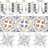
Figure 1.. Generation of hERG channel models in the closed (a), open (b), and inactivated (c) states.
The lower limit of the pore radius color profile (1.15 Å) indicates the minimum radius to accommodate a water molecule, and the upper limit (2.30 Å) indicates sufficient space to fit two water molecules side-by-side. ‘Max seq’ is a setting in ColabFold that denotes the maximum number of cluster centers and extra sequences that the multiple sequence alignment (MSA) used for AlphaFold2 will be subsampled to. ‘# models’ indicates the number of models predicted using the provided structural templates.
Figure 1—figure supplement 1.
Figure 1—figure supplement 1.. An example illustrating the extension of our strategy to model alternative ion channel states using AlphaFold.
Deactivated VSD II from the cryo-EM structure of NaV1.7 (PDB 6N4R) (Xu et al., 2019) is isolated and used as a structural template input for AlphaFold with a subsampled MSA. This guides AlphaFold to generate an NaV1.5 model with VSDs in the deactivated (S4 helices downward) conformation, representing a plausible closed state. Compared to a cryo-EM open-state NaV1.5 structure (PDB 6LQA, rightmost) (Li et al., 2021a) with activated VSDs (S4 helices upward), the AlphaFold-predicted model displays a visibly narrower pore. This example demonstrates how our approach using targeted structural biasing can be used to model different ion channel states beyond hERG.
Figure 2.
Figure 2.. Clustering of AlphaFold2-predicted hERG channel models.
(a) Clusters created from 100 models predicted for each state. Each structure visualized is colored according to the per-residue confidence metric (predicted Local Distance Difference Test, pLDDT). The closed-state models are clustered based on the backbone Cα RMSD of the entire protein models. The inactivated and open-state models are clustered based on the all-atom RMSD of the selectivity filter (residues S624–G628). To represent each cluster, the top 5 models ranked by an average pLDDT are shown. The bar graphs display the mean pLDDT values for the clustered segments across all models within each cluster, with the standard deviations shown as error bars. Clusters containing less than three models are categorized as outliers. (b) The models chosen for subsequent analysis colored by per-residue pLDDT values.
Figure 2—figure supplement 1.
Figure 2—figure supplement 1.. Protein backbone dihedral angle distributions of AlphaFold predicted models during inactivated-state-sampling and reference structures across key residues involved in ion selectivity and drug binding.
Histograms show the distribution of Phi (φ) and Psi (ψ) protein backbone dihedral angles across 100 AlphaFold-predicted models and associated subunits (four each) for residues in the selectivity filter (S624 – G628) and drug-binding site residues in the pore-lining S6 segment (Y652 and F656). Overlayed colored markers represent φ or ψ dihedral angles from the reference models used in the study: Open (PDB 5VA2, blue circles), AlphaFold inactivated-state-sampling representative models from cluster 2 and 3 (orange squares and purple diamonds, respectively), and closed (green triangles). The marker positions, representing the φ or ψ dihedral angles of the key residues from all four subunits of the reference models, are placed along the x-axis at their corresponding angle values. Their height on the y-axis does not reflect how often they occur, but it is simply adjusted to prevent the markers from overlapping with each other. These distributions reveal that reference models span the dominant conformational populations of selectivity filter and drug binding residues predicted by AlphaFold. Since the reference models were relaxed using Rosetta to resolve steric clashes and improve structural quality, some of these marker angles may deviate slightly from the populated regions in the histograms.
Figure 2—figure supplement 2.
Figure 2—figure supplement 2.. Comparison of the SF in hERG closed- (a, d), open- (b, e), and inactivated-state (c, f) models.
(a-c) Measurement of the distances between each carbonyl oxygen lining the conduction pathway in the SF. In the open- and closed-state models, S620 backbone carbonyl interacts with G626 and S624 backbone amide NH groups. In the inactivated-state model, the hydrogen bond between S620 and G626 is absent due to a reorientation of V625 backbone. However, at the bottom of the SF, S624 sidechain interacts with S623 backbone carbonyl from an adjacent subunit (denoted by *). (d-f) View of the SF from the extracellular side. For the closed- and inactivated-state models large black arrows indicate the rotation of the F627 side chains, while small black arrows show the rotation of the loops that connect the upper SF to the S6 helix, all relative to the equivalent structural elements in the open-state model.
Figure 3.
Figure 3.. Structural comparison of different hERG channel state models.
(a) Visual comparison of the closed-, open-, and inactivated-state models. (b) Pore radius for the selectivity filter (SF) and drug-binding region (upper) and for the entire pore (lower). (c) Comparison of the voltage sensing domain (VSD) conformation in each model, showcasing the positively charged Arg and Lys gating-charge residues (yellow), located on the S4 helix, and the gating charge transfer center residue, F463 (magenta), on the S2 helix. (d) Distances between the Cα atom of residue F463 to the Cα atom of each of the gating-charge residues.
Figure 3—figure supplement 1.
Figure 3—figure supplement 1.. Distance-based contact maps comparing intra- and intersubunit contacts between each model.
Two residues whose Cα atoms are within 6 Å of each other are considered to be in contact, provided there are no Cα atoms belonging to a third residue in between. Black cells indicate no contacts. Gray cells indicate a contact is present in both states being compared. Blue, orange, and green colored cells indicate the interaction is present only in the open, inactivated, or closed state, respectively, but not in the other state being compared in the map. Colored topology labels are included along the left and bottom edges of the maps showing the specific segments of the hERG channel to which the residues correspond.
Figure 3—figure supplement 2.
Figure 3—figure supplement 2.. Comparison of the S6 helix conformation for the hERG closed- (a), open- (b), and inactivated-state (c) models.
Residues E575 – L666 from the pore domain are visualized as dark gray ribbons. Selectivity filter (SF) residues and those on the S6 helix are shown with their backbone and side chains displayed as colored sticks. C atoms are gray, O are red, N are blue, S are yellow, H are not shown. The drug binding residues Y652 and F656 are highlighted in green.
Figure 4.
Figure 4.. Interaction network analysis showcasing residue–residue interactions in the S5-P linker (residues I583–Q592) and region surrounding the selectivity filter (SF) (residues S620–N633).
(a) An image of a hERG channel subunit with the analyzed S5-P linker and SF regions colored in light green and light blue, respectively. (b) Heatmaps showing intrasubunit and intersubunit (marked by X) interactions between each residue in the analyzed regions. The interactions analyzed are hydrogen bonding, π stacking, cation–π, and salt bridges. Black cells indicate no interactions. Gray cells indicate an interaction is present in both states. Blue, orange, and green colored cells indicate the interaction is present only in the open, inactivated, or closed state, respectively, but not in the other state being compared in the map. White lines are added to separate S5-P linker residues from the SF region residues. (c–e) Visualization of the interactions being present in one state but not the other. Gold-colored residues are involved in the interactions. Green-colored residues, named with an asterisk at the end, are from an adjacent subunit but are interacting with gold-colored residues. Dashed lines represent hydrogen bonds.
Figure 5.
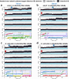
Figure 5.. Movement of K+ ions through hERG selectivity filter (SF) during all-atom molecular dynamics (MD) simulations with the applied membrane voltage.
The z coordinates of K+ ions are tracked as they traverse the pore of the hERG channel from the intracellular gate (lower y-axis limit) to the extracellular space (upper y-axis limit) under the membrane voltage of 750 mV. Putative K+-binding sites in the SF (S0–S5) are marked using blue dashed lines in the plots. Results from MD simulations on the open-state model with the SF occupancy initially configured to have only K+ ions (a) or alternating K+/water molecules (c), respectively. Results from MD simulations on the inactivated model with the SF occupancy initially configured to have only K+ ions (b) or alternating K+/water molecules (d), respectively.
Figure 5—figure supplement 1.
Figure 5—figure supplement 1.. Setup of MD simulations to assess ion conduction in the open and inactivated hERG channel models.
(a) Initial configuration of the SF, set to fill with either all K+ ions (top), or alternating K+ and water molecules (bottom). (b) An example MD simulation box showing a hERG channel model (shown in yellow surface representation) embedded in POPC lipid bilayer (shown as sticks) and solvated by an aqueous 0.3 M KCl solution (shown as a transparent surface with K+ and Cl- ions shown as purple and green balls, respectively).
Figure 5—figure supplement 2.
Figure 5—figure supplement 2.. Movement of K+ ions through hERG selectivity filter (SF).
The z coordinates of K+ ions are tracked as they traverse through the pore of the channel from the intracellular gate (lower y-axis limit) to the extracellular space (upper y-axis limit). Putative K+ binding sites in the SF (S0 – S5) are marked using blue dashed lines in the plots. (a, c) Molecular dynamics (MD) simulations with the applied 500 mV membrane voltage of the open-state model with the SF initially configured to have only K+ ions (a) or alternating K+ / water molecules (c), respectively. (b, d) MD simulations with the applied 500 mV membrane voltage of the inactivated-state model with the SF initially configured to have only K+ ions (b) or alternating K+ / water molecules (d), respectively. (e, g) MD simulations without applied membrane voltage of the open-state model with the SF initially configured to have only K+ ions (e) or alternating K+ / water molecules (g), respectively. (f, h) MD simulations without applied membrane voltage of the inactivated-state model with the SF initially configured to have only K+ ions (f) or alternating K+ / water molecules (h), respectively.
Figure 5—figure supplement 3.
Figure 5—figure supplement 3.. Analysis of modulations of the selectivity filter (SF) conformations and pore radii over the course of the 1 µs long molecular dynamics (MD) simulations.
The blue/orange-colored lines represent the average pore radii, and the shaded regions represent the standard deviation measured in MD simulations for a given Z value. The black lines represent the initial pore radii. The label on the left indicates the voltage of the MD simulations in each row.
Figure 5—figure supplement 4.
Figure 5—figure supplement 4.. Analysis of dynamics of the SF and pore conformations over the course of the 1 µs MD simulations.
(a) Pore radius averaged over each 1 µs long MD simulations with (right) or without (left) applied membrane voltage. Open- and inactivated-state model MD simulations are notated as O and I, respectively, with the subscripts KK and WK denoting whether the SF initially configured to have only K+ ions or alternating K+ / water molecules, respectively. (b) Ensembles of SF conformation over the course of each MD simulation superimposed. The golden-colored conformation indicates the initial conformation.
Figure 5—figure supplement 5.
Figure 5—figure supplement 5.. Representative model from the AlphaFold predicted inactivated-state-sampling cluster 3 (AF ic3).
(a) Structural overview of the representative model from AlphaFold inactivated-state sampling cluster 3, with pore radius mapped along the ion conduction pathway. The SF is highlighted, showing a flipped G626 carbonyl oxygen, deviating from the canonical ion-coordination geometry. (b) Structural comparison of the cluster 3 model with closed, open, and inactivated state models. Despite the G626 rearrangement, the overall pore conformation most closely resembles the open-state structure. (c-d) Molecular dynamics simulations under 750 mV applied voltage show K⁺ ion permeation through the inactivated-state-sampling cluster 3 model. The z-coordinates of K⁺ ions are plotted over time, tracking their position along the pore axis from intracellular (bottom) to extracellular (top) side. Horizontal dashed lines indicate canonical K⁺ binding sites in the selectivity filter (S0–S5). (c) MD simulation with K⁺ ions initially placed in the selectivity filter. (d) MD simulation with both K⁺ ions and water molecules initially placed in the selectivity filter. In both cases, K⁺ ions stably occupied and permeated the SF across on the microsecond timescales, demonstrating that this model supports K⁺ conduction. These results suggest that, despite structural deviations from the PDB 5VA2-based open-state model, the cluster 3 model represents an alternative open-like conformation functionally capable of ion conduction.
Figure 5—figure supplement 6.
Figure 5—figure supplement 6.. Cross-subunit distances between carbonyl oxygens of open-state hERG selectivity filter residues during MD simulations under different applied voltage and initial K+ ion position conditions.
Movement of potassium ions (denoted by differently colored lines) across the SF is shown at the bottom for reference. In top graphs red lines indicate initial distances. Labels 1 and 2 in red and blue, respectively, indicate a sequential dilation process exhibited by the hERG channel: the SF near residues F627 dilates first, followed by that around G628 SF residues.
Figure 5—figure supplement 7.
Figure 5—figure supplement 7.. Sequential dilation steps of hERG upper selectivity filter (SF).
SF residues are shown as gray sticks, water molecules as red and white spheres, and K+ as purple spheres. The first step, occurring around 100 ns, involves the flipping of F627 carbonyl oxygen, creating a small dilation at this level. At 500 ns, further dilation can be seen at the level of residues F627 and G628 in one subunit. At 1000 ns, the entire upper region of the SF dilates further. Frames were taken from an MD simulation of the open-state hERG channel with K+ and water initially in the SF prior to application of the transmembrane voltage of 750 mV.
Figure 6.
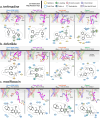
Figure 6.. Visualization of interactions for terfenadine (a), dofetilide (b), and moxifloxacin (c) with different hERG channel models.
Each panel includes four subpanels showcasing drug interactions with the open- (PDB 5VA2-derived and AlphaFold-predicted from inactivated-state-sampling Cluster 3, i.e., AF ic3), inactivated-, and closed-state hERG channel models. The estimated drug-binding free energies, ΔGbind, are given in Rosetta energy units (R.E.U.) and shown as averages ± standard deviations. In each subpanel, an overview of where the drug binds within the hERG channel pore is shown on the upper left, a 3D visualization of interactions between each channel residue (blue, red, green, and tan colored residues are from the subunit A, B, C, or D, respectively) to the drug (magenta) is shown on the upper right, and a 2D ligand–protein interaction map is shown at the bottom. A continuous gray line depicts the contour of the protein-binding site, and any breaks in this line indicate areas where the ligand is exposed to the solvent.
Figure 6—figure supplement 1.
Figure 6—figure supplement 1.. GALigandDock drug docking free energies for different hERG channel models.
Each bar plot displays the estimated binding free energy (Rosetta Energy Units, R.E.U.) for the specified drug across hERG channel models: Open (PDB 5VA2-based, blue), Open (AlphaFold inactivated-state-sampling Cluster 3, AFic3, purple), Inactivated (AlphaFold inactivated-state-sampling Cluster 2, orange), and Closed (green) states, with lower values indicating more favorable binding. For each drug and hERG channel model, 25,000 docking poses were generated, and the top 100 lowest-energy poses were clustered. The plotted values represent the mean and standard deviation of the best cluster, selected using a hybrid scoring method that considers both binding free energy and cluster size, with preference given to non-outlier clusters within a defined ΔG tolerance of 0.25 R.E.U. Suffixes (0), (+), and (±) denote the drug’s neutral, cationic, or zwitterionic form, respectively. Additional suffixes indicate experimental validation (Alexandrou et al., 2006; Duan et al., 2007; Numaguchi et al., 2000; Perrin et al., 2008; Suessbrich et al., 1997; Wang et al., 1997) for preferential binding to the inactivated state (*) or no preference (†).
Figure 6—figure supplement 2.
Figure 6—figure supplement 2.. Astemizole (a) and E-4031 (b) binding to different hERG channel models and cryo-EM structures.
Each panel includes 4 subpanels showcasing drug interactions with the open- (PDB 5VA2-derived and AlphaFold-predicted from inactivated-state-sampling cluster 3 i.e., AF ic3) and inactivated hERG channel models as well as the cryo-EM drug-bound hERG channel structures (PDB IDs 8ZYO and 8ZYP for astemizole and E-4031, respectively). The estimated drug binding free energies, ΔGbind, are given in Rosetta energy units (R.E.U) and shown as averages ± standard deviations. In each subpanel, an overview of where the drug binds within the hERG channel pore is shown on the upper left, a 3D visualization of interactions between each hERG channel residue (blue, red, green, and tan colored residues are from the subunit A, B, C, or D, respectively) to the drug (magenta) is shown on the upper right, and a 2D ligand – protein interaction map is shown at the bottom. A continuous gray line depicts the contour of the protein binding site, and any breaks in this line indicate areas where the ligand is exposed to the solvent.
Figure 7.
Figure 7.. Correlation of simulated hERG drug-binding affinities with experimental drug potencies under different modeling scenarios.
Single- and multi-state simulated drug-binding affinities (in Rosetta energy units, R.E.U.) are plotted against experimental drug potencies (IC₅₀ converted to free energies in kcal/mol). Lower (more negative) values indicate stronger binding. Horizontal error bars reflect uncertainty from experimental IC50 measurements, while vertical bars reflect standard deviations in simulated drug-binding affinities (n = 100), propagated across ionization states and channel state distributions. A total of 23 measurements representing 16 unique drugs were analyzed. Linear regression was performed using the least-squares method, and exact values for Pearson’s r, R2, and p-values are reported within the figure. (a) Single-state docking using the experimentally derived open-state structure (PDB 5VA2) yields a moderate correlation (the coefficient of determination R2 = 0.43, Pearson correlation coefficient r = 0.66). (b) Multi-state docking incorporating open (PDB 5VA2), inactivated, and closed-state conformations weighted by experimentally observed state distributions further improve the correlation (R2 = 0.63, r = 0.79). (c) Single-state docking using an alternative AlphaFold-predicted open state (inactivated-state-sampling Cluster 3, ic3) (R2 = 0.44, r = 0.66). (d) Multi-state docking combining the AlphaFold-predicted open- (inactivated-state-sampling Cluster 3, ic3), inactivated-, and closed-state models also results in a notable improvement (R2 = 0.64, r = 0.80) compared to single-state docking in panel (c) and comparable performance to multi-state docking in panel (b). These results highlight the enhanced predictive power of multi-state modeling and suggest that structural diversity within ensembles can compensate for individual model limitations, yielding more accurate predictions of drug–ion channel interactions and their effect on ion channel function.

Update of

Similar articles

Cited by

References

    1. Abi-Gerges N, Holkham H, Jones EMC, Pollard CE, Valentin JP, Robertson GA. hERG subunit composition determines differential drug sensitivity. British Journal of Pharmacology. 2011;164:419–432. doi: 10.1111/j.1476-5381.2011.01378.x. - DOI - PMC - PubMed
    1. Abramson J, Adler J, Dunger J, Evans R, Green T, Pritzel A, Ronneberger O, Willmore L, Ballard AJ, Bambrick J, Bodenstein SW, Evans DA, Hung C-C, O’Neill M, Reiman D, Tunyasuvunakool K, Wu Z, Žemgulytė A, Arvaniti E, Beattie C, Bertolli O, Bridgland A, Cherepanov A, Congreve M, Cowen-Rivers AI, Cowie A, Figurnov M, Fuchs FB, Gladman H, Jain R, Khan YA, Low CMR, Perlin K, Potapenko A, Savy P, Singh S, Stecula A, Thillaisundaram A, Tong C, Yakneen S, Zhong ED, Zielinski M, Žídek A, Bapst V, Kohli P, Jaderberg M, Hassabis D, Jumper JM. Accurate structure prediction of biomolecular interactions with AlphaFold 3. Nature. 2024;630:493–500. doi: 10.1038/s41586-024-07487-w. - DOI - PMC - PubMed
    1. Alexandrou AJ, Duncan RS, Sullivan A, Hancox JC, Leishman DJ, Witchel HJ, Leaney JL. Mechanism of hERG K+ channel blockade by the fluoroquinolone antibiotic moxifloxacin. British Journal of Pharmacology. 2006;147:905–916. doi: 10.1038/sj.bjp.0706678. - DOI - PMC - PubMed
    1. AlQuraishi M. AlphaFold at CASP13. Bioinformatics. 2019;35:4862–4865. doi: 10.1093/bioinformatics/btz422. - DOI - PMC - PubMed
    1. Asai T, Adachi N, Moriya T, Oki H, Maru T, Kawasaki M, Suzuki K, Chen S, Ishii R, Yonemori K, Igaki S, Yasuda S, Ogasawara S, Senda T, Murata T. Cryo-EM Structure of K+-Bound hERG channel complexed with the Blocker Astemizole. Structure. 2021;29:203–212. doi: 10.1016/j.str.2020.12.007. - DOI - PubMed

MeSH terms

Substances