. 2025 Jul 14;163(1):76.
doi: 10.1007/s00418-025-02400-6.

Differentiation to insulin-positive cells from human amnion epithelial cells using a pancreatic development mimicry protocol

Affiliations

Differentiation to insulin-positive cells from human amnion epithelial cells using a pancreatic development mimicry protocol

Daniel Martínez-Rodríguez et al. Histochem Cell Biol. .

Abstract

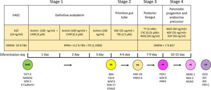
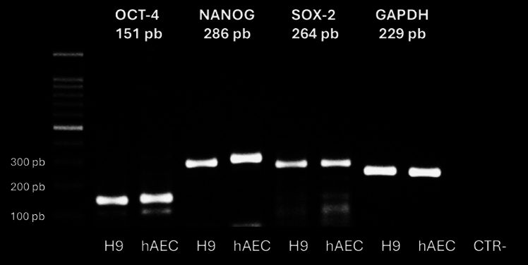
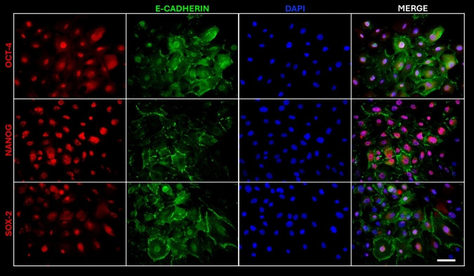
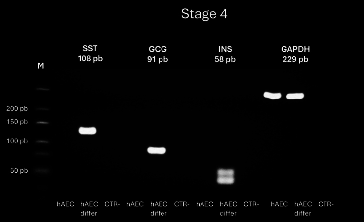
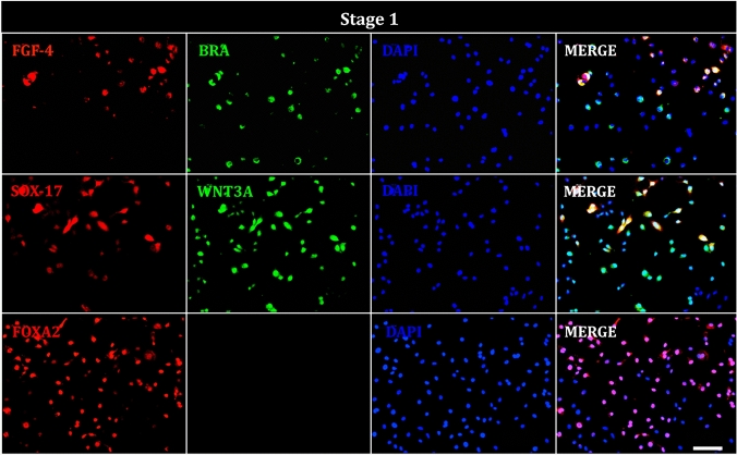
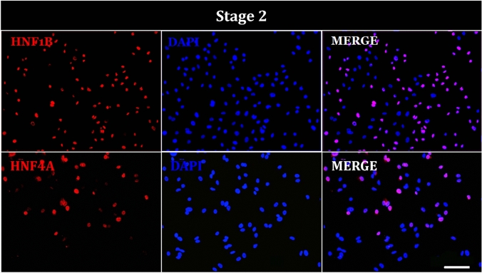
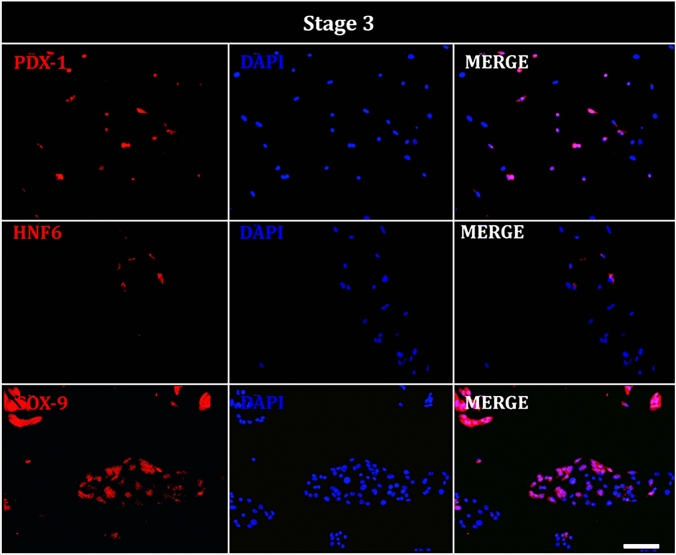
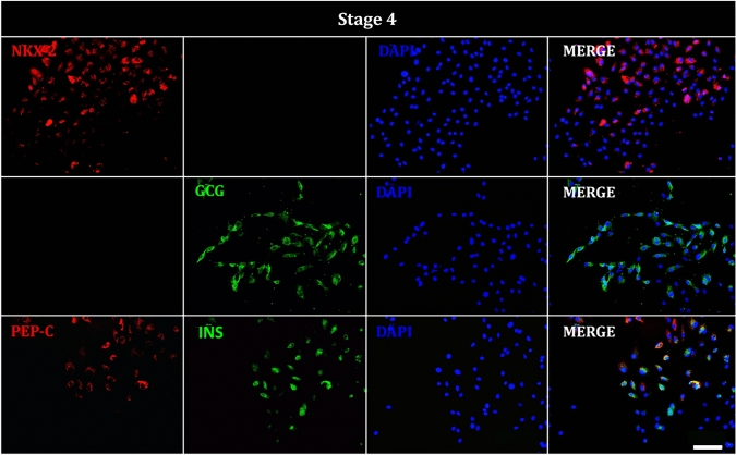
Diabetes mellitus (DM) is characterized by the loss or dysfunction pancreatic β-cells. Human amniotic epithelial cells (hAEC), which retain pluripotency markers and are readily obtainable from term placentas, represent a promising alternative source of stem cells. We investigated whether hAECs can be guided through pancreatic ontogeny to generate insulin-producing β-like cells. hAEC from uncomplicated term deliveries were expanded to passage 1 and exposed to a four-stage differentiation sequence that sequentially modulated Activin/WNT, KGF/TGF-β, retinoic-acid/hedgehog, and EGF/Noggin signaling. Stage progression was monitored by end-point RT-PCR and quantitative immunofluorescence for hallmark transcription factors. After definitive endoderm induction, 64% of cells were Brachyury positive and 71% were WNT3A positive; primitive-gut specification yielded 57% HNF1B+ cells. Posterior foregut commitment produced 75% PDX1+ and 60% Sox9+ nuclei and the final endocrine stage generated 74% NKX2.2+ cells, with 71% showing cytoplasmatic insulin and 51% C-peptide staining; insulin/C-peptide co-localization was confirmed by double labelling. Thus a chemically defined, four-step protocol can convert term-derived hAEC into insulin-producing β-like cells, supporting their use as an accessible platform for diabetes modelling and future cell-replacement therapies.

Keywords: Differentiation; Human amniotic epithelial cells; Pancreatic beta cells.


Conflict of interest statement

Declarations. Conflict of interest: The authors declare no competing interests.

Figures

Fig. 1
Fig. 1
Schematic of four-stage differentiation protocol from human amniotic epithelial cells (hAEC) into definitive endoderm, primitive gut tube, posterior foregut, and mixed population comprising pancreatic endoderm and endocrine precursor cells
Fig. 2
Fig. 2
Reverse transcription polymerase chain reaction (RT-PCR) analysis of pluripotency marker gene expression in hAEC, including OCT-4, NANOG, and SOX-2. Positive control; human embryonic stem cell line H9 (CTR+). Negative control (CTR−): reactions without cDNA
Fig. 3
Fig. 3
Microphotographs of hAEC positive for pluripotency markers OCT-4, NANOG, and SOX-2 (red), E-cadherin (green) and DAPI (blue) staining the nuclei. Image taken at ×20. Scale bar = 100 μm
Fig. 4
Fig. 4
RT-PCR analysis of gene expression in hAEC at the end of stage 1 of differentiation. Expression of constitutive gene glyceraldehyde-3-phosphate dehydrogenase (GAPDH) in undifferentiated hAEC and hAEC at the end of the protocol (differ). The gels show the RT-PCR products for BRACHYURY (122 bp), SOX17 (151 bp), and GADPH (229 bp). Negative control (CTR): reactions without cDNA
Fig. 5
Fig. 5
RT-PCR analysis of gene expression in hAEC at the end of stage 4 of differentiation protocol into insulin-positive cells. Expression of somatostatin (SST), glucagon (GCG), insulin (INS) and the constitutive gene glyceraldehyde-3-phosphate dehydrogenase (GAPDH) in undifferentiated and differentiated (differ) hAEC. The gels shown RT-PCR products for SST (108 bp), CGC (91 bp), INS (58 bp), and GAPDH (229 bp). Negative control (CTR): reactions without cDNA
Fig. 6
Fig. 6
Immunofluorescence of hAEC at stage 1 of differentiation. Representative images show cells positive for definitive endoderm markers; FGF-4 (red), BRACHYURY (BRA, green), SOX-17 (red), WNT3A (green), FOXA2 (red), and DAPI (blue) staining the nuclei. Image taken at ×20. Scale bar = 100 μm
Fig. 7
Fig. 7
Immunofluorescence of hAEC at stage 2 of differentiation. Representative images show cells positive for primitive gut tube markers; HNF1B (red), HNF4A (red), and DAPI (blue) staining the nuclei. Image taken at ×20. Scale bar = 100 μm
Fig. 8
Fig. 8
Immunofluorescence of hAEC at stage 3 of differentiation. Representative images show cells positive for posterior foregut markers; PDX-1 (red), HNF-6 (red), SOX-9 (red), and DAPI (blue) staining the nuclei. Image taken at ×20. Scale bar = 100 μm
Fig. 9
Fig. 9
Immunofluorescence of hAEC at stage 4 of differentiation. Representative images show cells positive for pancreatic progenitor and endocrine precursor markers; NKX2 (red), glucagon, (GCG, green), C-peptide (C-PEP, red), insulin (INS, green), and DAPI (blue) staining the nuclei. Image taken at ×20. Scale bar = 100 μm
Fig. 10
Fig. 10
Quantification of marker-positive cells at each stage of the differentiation protocol from human amniotic epithelial cells (hAEC)

Similar articles

References

    1. Abouna GM (2008) Organ shortage crisis: problems and possible solutions. Transplant Proc 40(1):34–38. 10.1016/j.transproceed.2007.11.067 - PubMed
    1. Agulnick AD, Ambruzs DM, Moorman MA, Bhoumik A, Cesario RM, Payne JK, Kelly JR, Haakmeester C, Srijemac R, Wilson AZ, Kerr J, Frazier MA, Kroon EJ, D’Amour KA (2015) Insulin-producing endocrine cells differentiated in vitro from human embryonic stem cells function in macroencapsulation devices in vivo. Stem Cells Transl Med 4(10):1214–1222. 10.5966/sctm.2015-0079 - PMC - PubMed
    1. American Diabetes Association (2015) (7) Approaches to glycemic treatment. Diabetes Care 38(Suppl):S41-48. http://doi.org/10.2337/dc15-S010 - PubMed
    1. American Diabetes Association Professional Practice Committee (2024a) 2 Diagnosis and classification of diabetes: standards of care in diabetes-2024. Diabetes Care 47(Supp1):S20–S42. 10.2337/dc24-S002 - PMC - PubMed
    1. American Diabetes Association Professional Practice Committee (2024b) 9. Pharmacologic approaches to glycemic treatment: standards of care in diabetes-2024. Diabetes Care 47(1):S158–S178. 10.2337/dc24-S009 - PMC - PubMed