. 2025 Jul 13;13(7):e010898.
doi: 10.1136/jitc-2024-010898.

Unveiling the mechanisms of CEBPD-orchestrated TAM polarization through RGS2/PAR2 pathway and its impact on ICB efficacy in clear cell renal cell carcinoma

Affiliations

Unveiling the mechanisms of CEBPD-orchestrated TAM polarization through RGS2/PAR2 pathway and its impact on ICB efficacy in clear cell renal cell carcinoma

Xiu-Wu Pan et al. J Immunother Cancer. .

Abstract

Background: The polarization status and function of tumor-associated macrophages (TAMs) influence tumor progression and patients' prognosis. CCAAT/enhancer-binding proteins (CEBPs) family are important transcriptional factors in macrophages differentiation physically and pathologically. This study aims to explore the mechanism of CEBPs in TAMs polarization in clear cell renal cell carcinoma (ccRCC) immune microenvironment and its impact on immune checkpoint blockers (ICBs) therapy.

Methods: The expression of CEBPs in ccRCC was analyzed by single-cell transcriptome and western blot. Immunofluorescence and in-vitro polarization assay were used to evaluate the effect of CEBP delta (CEBPD) on TAMs. Chromatin immunoprecipitation sequencing was used to explore targets of CEBPD. Dual-luciferase reporter assay and electrophoretic mobility shift assay were performed to confirm the regulation of CEBPD to RGS2. Specimens of patients received ICB therapy were used to analyze the relationship between CEBPD and immunotherapy.

Results: The study identified CEBPD as a key transcriptional factor in ccRCC TAM polarization. Upregulation of CEBPD correlates to decreased M1/M2 ratio of TAMs and poorer clinical outcomes. CEBPD inhibited M1-like polarization in vitro and in vivo via the RGS2/PAR2 axis. Furthermore, CEBPD also affected the therapeutic efficacy of ICB.

Conclusion: This study revealed CEBPD regulated TAM polarization via the CEBPD/RGS2/PAR2 axis. Targeting CEBPD may be a potential approach and a complementary strategy to ICB therapies in ccRCC.

Keywords: Immunotherapy; Kidney Cancer; Macrophage.


Conflict of interest statement

Competing interests: None declared.

Figures

Figure 1
Figure 1. CEBPD is upregulated in ccRCCs and correlated with impaired immune response. (A) The cell type clusters of myeloid cells. (B) The group clusters of myeloid cells. (C) The proportion of myeloid cells in ccRCCs and adjacent kidney tissues. (D) The expression of CEBPD family in cell type clusters, and the yellow dot represents high level of average expression, while the purple dot represents low level of average expression. Besides, the size of the dot represents the percentage of cells expressing the gene. Transcription factor activity of (E) CEBPB and (F) CEBPD in myeloid cells. (G) Proportion of 5 groups of myeloid cells in CEBPD-high and CEBPD-low ccRCCs. (H) Pathway enrichment analysis of 13 ccRCC samples. (I) Heatmap of cytokines expression of 13 ccRCC samples. (J) Volcano map of differentially expressed genes between CEBPD-high and low TAM3. Data were presented as mean±SD. A significant difference between the groups, **p<0.01, ***p<0.001, and ****p<0.0001. ccRCC, clear cell renal cell carcinoma; CEBP, CCAAT/enhancer-binding protein; CEBPD, CEBP delta; DC, dendritic cell; GSVA: gene set variation analysis; IL, interleukin; ns, not significant; RCC, renal cell carcinoma; TAM, tumor-associated macrophage.
Figure 2
Figure 2. CEBPD upregulation influenced TAM proportion and was associated with poorer prognosis. (A) Western blot results showed the protein expression of CEBPA, CEBPB, CEBPD, CEBPG, CEBPZ in six patient specimens. (B) Kaplan-Meier curves of TCGA-KIRC cohort. (C) Kaplan-Meier curves of survival probability in Xinhua Hospital cohort. Patients were divided by IHC score. (D) Representative immunofluorescence images of adjacent kidney tissue (left), CEBPD-low tumor (middle) and CEBPD-high tumor (right), and (E) quantitative analysis of M1-like and M2-like TAMs.Data were presented as mean±SD. A significant difference between the groups, *p<0.05. ccRCC, clear cell renal cell carcinoma; CEBP, CCAAT/enhancer-binding protein; CEBPD, CEBP delta; IHC, immunohistochemistry; KIRC, kidney renal clear cell carcinoma; TAM, tumor-associated macrophage; TCGA, The Cancer Genome Atlas.
Figure 3
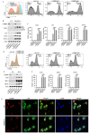
Figure 3. CEBPD knockdown enhanced M1 type polarization of THP-1 cells. (A) CD86 positive cells in CEBPD-kd1, CEBPD-kd2 and control group. (B–C) CEBPD knockdown enhanced the expression of IL-1β and IL-6. (D) CD206 positive cells in CEBPD-kd1, CEBPD-kd2 and control group. (E–F) CEBPD knockdown did not affect the expression of M2 markers. (G) CEBPD knockdown promoted the translocation of p65 from cytoplasm to nucleus. A significant difference between the groups, ***p<0.001. CEBPD, CCAAT/enhancer-binding protein delta; DAPI: 4',6-diamidino-2-phenylindole; IFN-γ, interferon-gamma; IL, interleukin; LPS, lipopolysaccharides; ns, not significant; UN, uninduced,
Figure 4
Figure 4. Chromatin immunoprecipitation sequencing analysis of CEBPD. (A) Pie chart of binding region of CEBPD. (B) Genomic distribution of CEBPD binding peaks. (C) Fold changes and (D) peak score of genes regulated by CEBPD. (E) Function enrichment of genes regulated by CEBPD. (F) Visualization of binding peak of CEBPD on promoter region of RGS2. Correlation analysis between CEBPD and RGS2 in (G) TAM3 and (H) TCGA-KIRC database. ccRCC, clear cell renal cell carcinoma; CEBPD, CCAAT/enhancer-binding protein delta; ChIP, chromatin immunoprecipitation; FC, fold change; KIRC, kidney renal clear cell carcinoma. TAM, tumor-associated macrophage; TCGA, The Cancer Genome Atlas.
Figure 5
Figure 5. CEBPD upregulated RGS2 by binding to its promoter region. (A) Dual-luciferase reporter assay results showed CEBPD bound to the first site on RGS2 promoter region. Western blot assay showed RGS2 increased as CEBPD was overexpressed while RGS2 decreased as CEBPD was knocked down in (B) THP-1 cells and (C) human monocytes. (D) EMSA results confirmed the combination between CEBPD protein and RGS2 promoter region. Data were presented as mean±SD. A significant difference between the groups, **p<0.01, ***p<0.001, and ****p<0.0001. BS, binding site. BS, binding site; BSA, bovine serum albumin; CEBPD, CCAAT/enhancer-binding protein delta; EMSA, electrophoretic mobility shift assay.
Figure 6
Figure 6. RGS2 inhibited M1 polarization through deactivating PAR2. (A) CEBPD knockdown increased IL-1β and IL-6, whereas RGS2 overexpression reversed this effect. (B) CEBPD knockdown promoted the translocation of p65 from cytoplasm to nucleus, whereas RGS2 overexpression reversed this effect. (C) PAR2 was upregulated during M1 polarization. (D) RGS2 knockdown activated PAR2-mediated MAPK signaling pathway. (E) PAR2 inhibitors reduced IL-1β and IL-6. (F) PAR2 inhibitors impeded the nuclear translocation of p65. CEBPD, CCAAT/enhancer-binding protein delta; DAPI: 4',6-diamidino-2-phenylindole; IFN-γ, interferon-gamma; IL, interleukin; LPS, lipopolysaccharides; PAR, proteinase-activated receptor; UN, uninduced.
Figure 7
Figure 7. Schematic diagram of CEBPD affecting M1-type polarization of macrophages through the RGS2/PAR2 axis. CEBPD, CCAAT/enhancer-binding protein delta; IL, interleukin; PAR, proteinase-activated receptor.
Figure 8
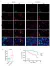
Figure 8. CEBPD correlated with ICB efficacy in patients with RCC. (A) Representative immunofluorescence images of patient respond to ICB therapy (left) and patient non-respond to ICB therapy (right), and (B) quantitative analysis of CEBPD intensity in two groups. (C) Kaplan-Meier curves of survival probability in CEBPD-high group and CEBPD-low group. CEBPD, CCAAT/enhancer-binding protein delta; ICB, immune checkpoint blocker; RCC, renal cell carcinoma.

References

    1. Hsieh JJ, Purdue MP, Signoretti S, et al. Renal cell carcinoma. Nat Rev Dis Primers. 2017;3:17009. doi: 10.1038/nrdp.2017.9. - DOI - PMC - PubMed
    1. Ljungberg B, Albiges L, Abu-Ghanem Y, et al. European Association of Urology Guidelines on Renal Cell Carcinoma: The 2022 Update. Eur Urol. 2022;82:399–410. doi: 10.1016/j.eururo.2022.03.006. - DOI - PubMed
    1. Choueiri TK, Motzer RJ. Systemic Therapy for Metastatic Renal-Cell Carcinoma. N Engl J Med. 2017;376:354–66. doi: 10.1056/NEJMra1601333. - DOI - PubMed
    1. Drake CG, Stein MN. The Immunobiology of Kidney Cancer. J Clin Oncol. 2018:JCO2018792648. doi: 10.1200/JCO.2018.79.2648. - DOI - PubMed
    1. The Cancer Genome Atlas Research Network Comprehensive molecular characterization of clear cell renal cell carcinoma. Nature New Biol. 2013;499:43–9. doi: 10.1038/nature12222. - DOI - PMC - PubMed

MeSH terms

LinkOut - more resources