. 2025 Jul 13;13(7):e011759.
doi: 10.1136/jitc-2025-011759.

T cell-specific non-viral DNA delivery and in vivo CAR-T generation using targeted lipid nanoparticles

Affiliations

T cell-specific non-viral DNA delivery and in vivo CAR-T generation using targeted lipid nanoparticles

Jaime Fernández Bimbo et al. J Immunother Cancer. .

Abstract

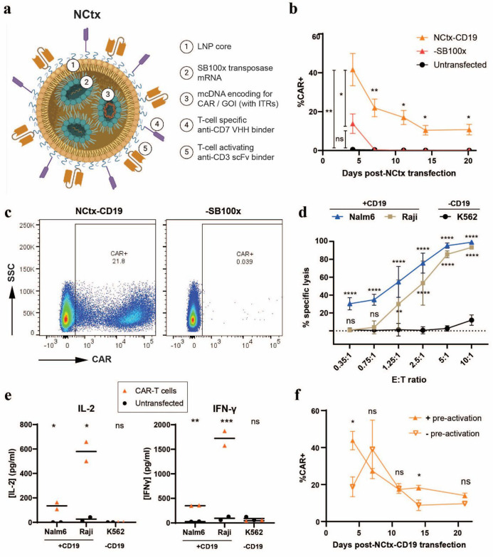
Background: Ex vivo chimeric antigen receptor (CAR)-T therapies have revolutionized cancer treatment. However, treatment accessibility is hindered by high costs, long manufacturing times, and the need for specialized centers and inpatient care. Strategies to generate CAR-T cells in vivo have emerged as a promising alternative that could bypass CAR-T manufacturing bottlenecks. Most current in vivo CAR-T approaches, while demonstrating encouraging preclinical efficacy, rely on transient messenger RNA (mRNA) delivery or viral vectors which both have limitations in terms of efficiency, durability, and scalability. To address these challenges, we developed a novel DNA-based targeted lipid nanoparticle (LNP) which we termed NCtx.

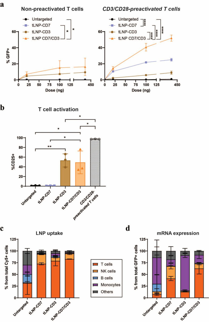
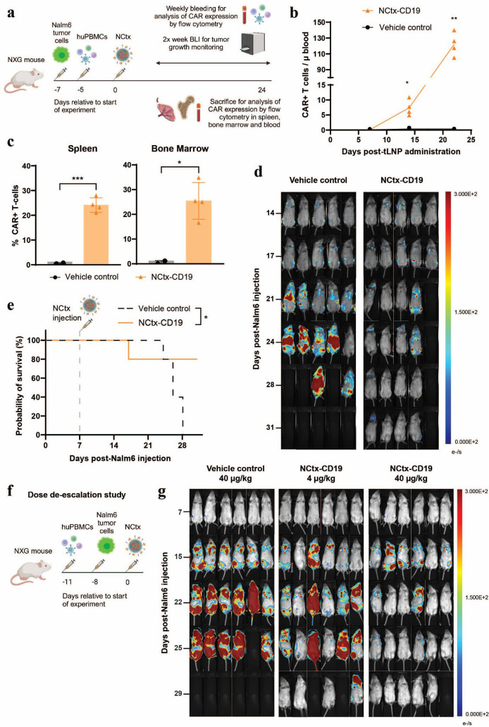
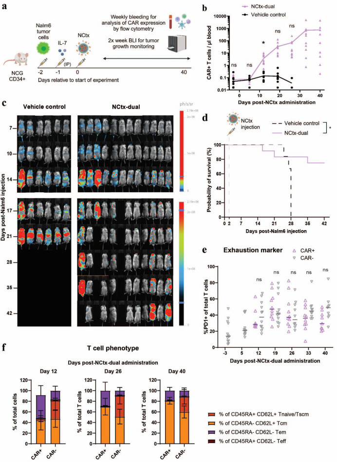
Methods: Minicircle DNA (mcDNA) encoding a CAR construct and SB100x transposase mRNA were encapsulated within a novel lipid formulation which was functionalized with T cell-specific anti-CD7 and anti-CD3 binders. In vitro, we evaluated T cell specificity, mcDNA and mRNA transfection efficiency, transposon-mediated CAR integration and functionality of the resulting CAR-T cells. In vivo efficacy was assessed in peripheral blood mononuclear cell and CD34+ stem cell humanized murine xenograft models of B cell leukemia.

Results: In vitro, NCtx displayed high specificity and transfection efficiency with both mcDNA and mRNA in primary T cells. Transposase mRNA facilitated genomic integration of the CAR gene, leading to the generation of stable CAR-T cells that exhibited antigen-specific cytotoxicity and cytokine release. In vivo, a single intravenous dose of NCtx induced robust CAR-T cell generation resulting in effective tumor control and significantly improved survival in two distinct xenograft models.

Conclusions: Our findings demonstrate for the first time that targeted LNPs can be employed for efficient DNA delivery to T cells in vitro and in vivo. We show that when combined with transposase technology, this LNP-based system can efficiently generate stable CAR-T cells directly in vivo, inducing potent and durable antitumor responses. NCtx represents a novel non-viral gene therapy vector for in vivo CAR-T therapy, offering a scalable and potentially more accessible alternative to traditional approaches in CAR-T cell generation.

Keywords: Chimeric antigen receptor - CAR; Gene therapy; Immunotherapy; Nanoparticle; T cell.


Conflict of interest statement

Competing interests: All authors are either full-time or part-time employees of NanoCell Therapeutics and hold equity or stock options in the company. MG, JL, EvD, DM, AA, ME, SN, NS and ZL are authors on patent applications related to this work. PM and MG are founders of Supercoiled GeneTx GmbH, which owns intellectual property rights to the minicircle DNA technology used in this study.

Figures

Figure 1
Figure 1. Dual-targeted anti-CD7/CD3 tLNPs functionally deliver mcDNA to T cells without exogenous activation while displaying high uptake and mRNA expression specificity. (a, b) Particles co-encapsulating reporter mCherry mRNA and eGFP mcDNA were either left unmodified (untargeted) or targeted with an anti-CD7 nanobody (tLNP-CD7), an anti-CD3 scFv (tLNP-CD3), or both (tLNP-CD7/CD3). These LNPs were applied to resting or CD3/CD28 bead preactivated T cells at a range of doses up to 400 ng total nucleic acid. (a) mcDNA expression after 96 hours shown as percentage eGFP+ cells measured by flow cytometry. (b) T cell activation status as measured by percentage of CD25+ cells by flow cytometry 96 hours post-treatment (400 ng dose). (a, b) n=3 T cell donors, data are mean±SD. (c, d) Surrogate particles co-encapsulating Cy5-labeled eGFP mRNA were applied to PBMCs at a dose of 20 ng to assess selectivity in a mixed cell population. (c) Uptake of the different LNP formulations was assessed after 2 hours as the percentage of each PBMC subset (T cells, NK cells, B cells, monocytes, and others) within the total Cy5+ population. (d) Functional mRNA delivery was evaluated after 24 hours as the percentage of each cell subset within the eGFP+ population. (d, e) n=2 T cell donors, data represent the mean±SD. P values were calculated using ordinary one-way ANOVA with Bonferroni correction for multiple comparisons. Where multiple doses were tested, statistics are calculated for the 100 ng dose. Only significant p values are plotted using *p<0.0332, **p<0.0021, ***p<0.0002 and ****p<0.0001. ANOVA, analysis of variance; LNP, lipid nanoparticle; mcDNA, minicircle DNA; mRNA, messenger RNA; NK, natural killer; PBMC, peripheral blood mononuclear cell; scFv, single-chain variable fragment; tLNP, targeted lipid nanoparticle.
Figure 2
Figure 2. NCtx-CD19 generates stable and functional CAR-T cells in vitro. (a) Schematic representation of NCtx, a targeted LNP platform for T cell engineering. The LNP (1) encapsulates mcDNA encoding a CAR or another GOI flanked by SB100x-compatible ITRs (2), along with SB100x transposase mRNA (3) to mediate genomic integration of the GOI upon transient expression of the transposase mRNA. The vehicle is functionalized with anti-CD7 VHH (4) and anti-CD3 scFv (5) targeting ligands. (b, c) CD3/CD28 bead-activated T cells were treated with a dose of 800 ng total nucleic acid using NCtx-CD19, surrogate particles lacking SB100x mRNA transposase (−SB100x) or left untreated (untransfected) for 4 days. Cells were monitored over 20 days to evaluate stable genomic integration. (b) Transfection efficiency as the percentage of CD19 CAR+T cells as measured by flow cytometry at various time points post-transfection. n=9 different T cell donors, data represent the mean±SD. (c) Representative flow plots at day 14 are shown. (d) Antigen-specific cytotoxicity of NCtx-CD19-engineered CAR-T cells, measured via luciferase-based killing assays against CD19+ (Nalm6, Raji) and CD19-negative (K562) target cells. CAR-T cells were collected 13 days post-transfection and co-cultured with target cells at varying effector-to-target (E:T) ratios for 24 hours. Specific lysis was calculated relative to untransfected control cells. n=9 using different T cell donors, data represent the mean±SD. (e) Antigen-specific cytokine release by NCtx-CD19-engineered CAR-T cells, measured as the secretion of proinflammatory cytokines IL-2 and IFN-γ. Cytokines were measured after a 24-hour co-culture of effector and target cells at an E:T ratio of 2.5:1, using both CAR+ and untransfected T cells. n=1, measured in technical duplicates. (f) Transfection efficiency of resting (− pre-activation) and bead-activated (+ pre-activation) T cells at various time points post-transfection as measured by flow cytometry analysis of CD19-CAR+cells. n=2 T cell donors in two independent experiments, data are presented as mean±SD. P values were calculated using two-way ANOVA, mixed effect model with Tukey correction for multiple comparisons (b, d, f) or unpaired t-test (e). Significance is plotted with ns for p>0.0322 and *p<0.0332, **p<0.0021, ***p<0.0002 and ****p<0.0001. In (d) Nalm6 and Raji (CD19+) are compared with K562 (CD19−). ANOVA, analysis of variance; CAR, chimeric antigen receptor; GOI, gene of interest; IFN-γ, interferon-gamma; IL-2, interleukin 2; ITR, inverted terminal repeat; LNP, lipid nanoparticle; mcDNA, minicircle DNA; mRNA, messenger RNA; ns, not significant; scFv, single-chain variable fragment; SSC, side scatter; VHH, variable domain of a heavy-chain-only antibody.
Figure 3
Figure 3. NCtx-CD19 generates CAR-T cells in vivo resulting in sustained tumor control across a range of doses in a human PBMC model of leukemia. (a) Schematic representation of study timeline: NXG mice were injected intravenously with 5×10⁵ luciferase-expressing Nalm6 tumor cells, followed by humanization with 5×10⁶ huPBMCs. NCtx-CD19 or a NCtx surrogate encapsulating eGFP mcDNA and SB100x mRNA (vehicle control) were administered intravenously at a total nucleic acid dose of 0.5 mg/kg. (b) Transfection efficiency in circulating T cells was measured over time via flow cytometry, represented as CAR+ cells/μL of blood. n=4 (NCtx-CD19) or n=5 (vehicle control), data are mean with individual values plotted. (c) CD19 CAR mcDNA expression was assessed by flow cytometry in spleen and bone marrow at the study’s end. Vehicle-treated mice reached HEP and were analyzed between days 24–28 (n=2), while NCtx-CD19-treated mice were all terminated and analyzed on day 31 (n=4). Data are presented as mean±SD. (d) Nalm6 tumor burden was monitored by BLI. (e) Kaplan-Meier survival analysis. Mouse death in the NCtx-CD19 group on day 24 post-Nalm6 was due to anesthetic overdose, unrelated to health or tumor burden. n=5. (f) Schematic representation of the dose de-escalation study timeline: NXG mice were humanized with 5×10⁶ huPBMCs 11 days pre-LNP injection, followed by intravenous injection with 5×10⁵ luciferase-expressing Nalm6 tumor cells 8 days before treatment. NCtx-CD19 or a NCtx vehicle control encapsulating only SB100x mRNA (vehicle control) were administered intravenously at a dose of total nucleic acid of either 40 or 4 µg/kg. (g) Nalm6 tumor burden was monitored via BLI. P values were calculated using two-way ANOVA, mixed effect model (b), unpaired t-test (c) or log-rank Mantel-Cox test (e). Significance is plotted *p<0.0332, **p<0.0021 and ***p<0.0002. ANOVA, analysis of variance; BLI, bioluminescent imaging; CAR, chimeric antigen receptor; HEP, humane endpoint; LNP, lipid nanoparticle; mcDNA, minicircle DNA; mRNA, messenger RNA; PBMC, peripheral blood mononuclear cell; tLNP, targeted lipid nanoparticle.
Figure 4
Figure 4. NCtx-dual induces robust and durable in vivo CAR-T generation, tumor control and extended survival in CD34+ HSC-engrafted NCG mice. (a) Schematic representation of study design: NCG mice engrafted with CD34+ HSC (NCG-His) were injected intravenously with 5×10⁵ luciferase-expressing Nalm6 tumor cells, followed by IP injection of 200 ng IL-7. Mice were treated intravenously with NCtx-dual or a NCtx vehicle control encapsulating eGFP mcDNA and SB100x mRNA (vehicle control) at a total nucleic acid dose of 50 µg/kg. (b) CD19/CD22 dual CAR mcDNA expression was assessed by flow cytometry in circulating T cells for 40 days post-NCtx administration. n=12, data are presented as mean with individual values. (c) Nalm6 tumor burden was monitored by BLI. (d) Kaplan-Meier survival analysis. n=6 (vehicle control) or n=12 (NCtx-dual). (e) Expression of the exhaustion marker PD-1 in CAR+ and CAR− T cell populations over time in NCtx-dual-treated mice, analyzed by flow cytometry. n=12, data represent mean±individual values. (f) T cell phenotype characterization (Tnaive/Tscm, Tcm, Tem, and Teff) based on CD45RA and CD62L expression in CAR+ and CAR− T cells after NCtx-dual administration. n=12, data represent mean±SD. P values were calculated using log-rank Mantel-Cox test (b) or two-way ANOVA, mixed effect model (d, e). Significance is plotted with ns for p>0.0332 and *p<0.0332. ANOVA, analysis of variance; BLI, bioluminescent imaging; CAR, chimeric antigen receptor; HSC, hematopoietic stem cell; IL-7, interleukin 7; IP, intraperitoneal; mcDNA, minicircle DNA; mRNA, messenger RNA; ns, not significant; PD-1, programmed cell death protein-1; Tcm, central memory T cell; Teff, effector T cell; Tem, effector memory T cell; Tnaive, naïve T cell; Tscm, stem cell memory T cell.

Similar articles

References

    1. Sengsayadeth S, Savani BN, Oluwole O, et al. Overview of approved CAR-T therapies, ongoing clinical trials, and its impact on clinical practice. EJHaem . 2022;3:6–10. doi: 10.1002/jha2.338. - DOI - PMC - PubMed
    1. Lu J, Jiang G. The journey of CAR-T therapy in hematological malignancies. Mol Cancer. 2022;21:194. doi: 10.1186/s12943-022-01663-0. - DOI - PMC - PubMed
    1. Vormittag P, Gunn R, Ghorashian S, et al. A guide to manufacturing CAR T cell therapies. Curr Opin Biotechnol. 2018;53:164–81. doi: 10.1016/j.copbio.2018.01.025. - DOI - PubMed
    1. Mikhael J, Fowler J, Shah N. Chimeric Antigen Receptor T-Cell Therapies: Barriers and Solutions to Access. JCO Oncol Pract . 2022;18:800–7. doi: 10.1200/OP.22.00315. - DOI - PubMed
    1. Ahmed N, Sun F, Teigland C, et al. Chimeric Antigen Receptor T-Cell Access in Patients with Relapsed/Refractory Large B-Cell Lymphoma: Association of Access with Social Determinants of Health and Travel Time to Treatment Centers. Transplantation and Cellular Therapy . 2024;30:714–25. doi: 10.1016/j.jtct.2024.04.017. - DOI - PubMed

LinkOut - more resources