. 2025 Jul 14;11(1):323.
doi: 10.1038/s41420-025-02556-z.

Hippo pathway and NLRP3-driven NETosis in macrophages: Mechanisms of viral pneumoniaaggravation

Affiliations

Hippo pathway and NLRP3-driven NETosis in macrophages: Mechanisms of viral pneumoniaaggravation

Bijun Luo et al. Cell Death Discov. .

Abstract

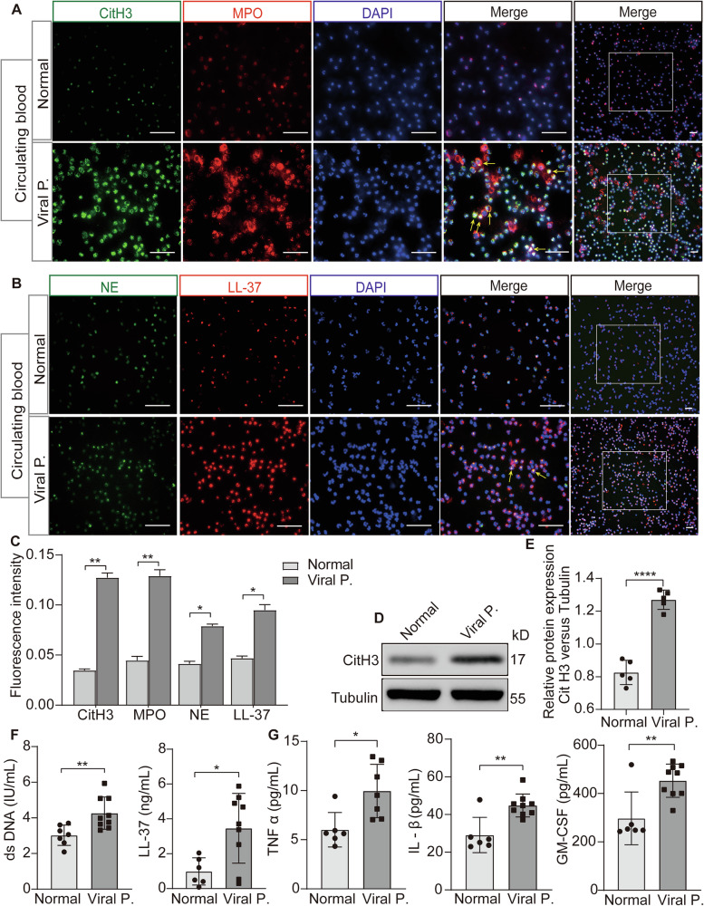
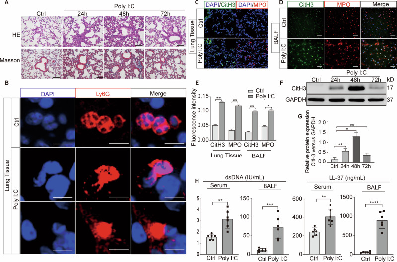
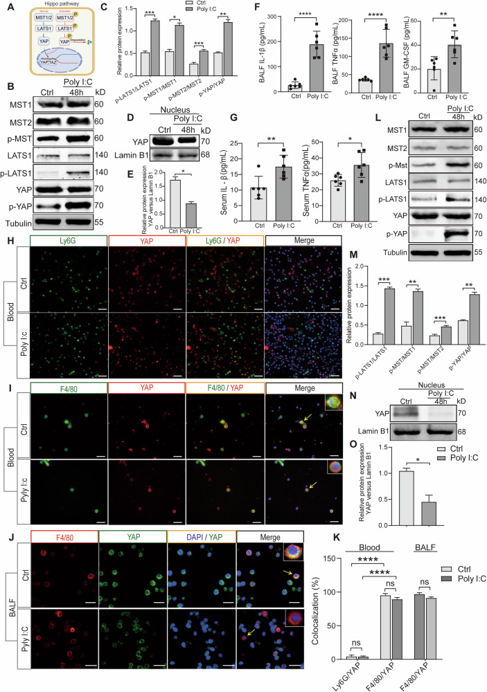
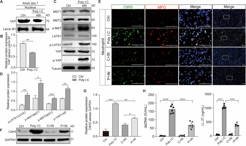
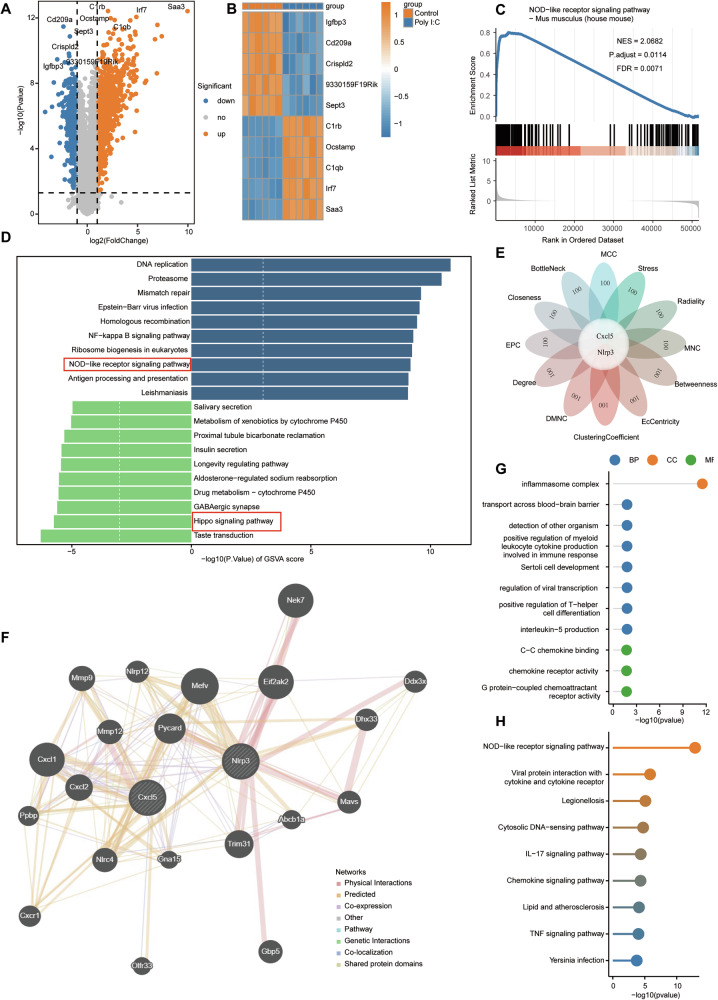
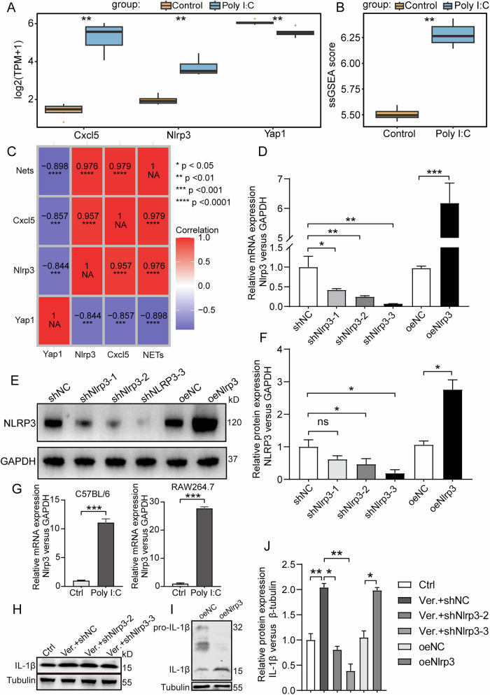
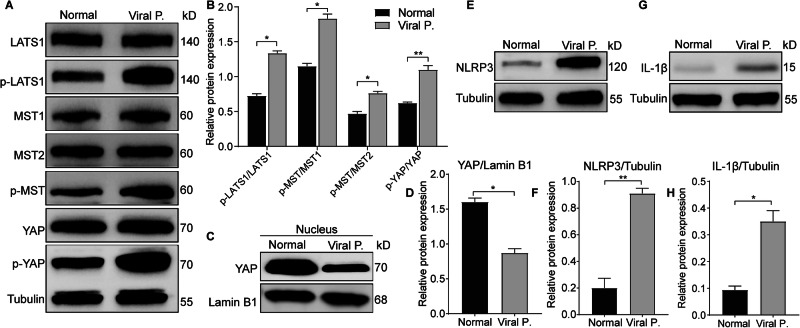
Severe viral infections can precipitate acute lung injury, resulting in significant morbidity and mortality. While NETosis serves as an important defense mechanism against pathogens and viruses, its excessive or dysregulated activation may contribute to pulmonary damage. In this study, elevated levels of NETosis were detected in the peripheral blood of patients with viral pneumonia. To further investigate the relationship between NETosis and virus-induced acute lung injury, a murine model was established using intratracheal administration of poly(I:C), a synthetic analog of double-stranded RNA that mimics viral infection. NETosis biomarkers were assessed in both patients and poly(I:C)-stimulated mice. In addition, we examined the role of the Hippo signaling pathway and its downstream mediators, including inflammatory factors and chemokines. Enhanced NETosis and activation of the Hippo pathway were observed in the lungs of poly(I:C)-treated mice, along with elevated levels of IL-1β in isolated macrophages. These effects were mitigated by Hippo pathway inhibitors. Co-culture experiments confirmed that IL-1β promotes NETosis, while NLRP3, acting downstream of the Hippo pathway, was responsible for IL-1β secretion. Patients with viral pneumonia showed increased NLRP3 and IL-1β expression in monocyte-derived macrophages compared to healthy controls. Overall, our findings indicate that activation of the Hippo pathway in macrophages during poly(I:C) exposure upregulates NLRP3 and IL-1β expression, thereby promoting NETosis and exacerbating virus-induced lung injury. This study highlights a potential therapeutic target to reduce lung damage caused by viral infections.


Conflict of interest statement

Competing interests: The authors declare that the research was conducted in the absence of any commercial or financial relationships that could be construed as a potential conflict of interest.

Figures

Fig. 1
Fig. 1. NETosis is involved in viral pneumonia with excessive inflammatory cytokines.
A, B Comparative analysis of NETosis biomarkers in neutrophils from peripheral blood of patients with viral pneumonia versus healthy volunteers via immunofluorescence. C Graphic presentations of fluorescence mean densities of NETosis biomarkers. D Assessment of CitH3 protein expression in whole blood sample from patients with viral pneumonia and healthy volunteers using western blotting. E Quantitative analysis of the protein CitH3 relative to Tubulin. F Quantification of serum dsDNA and LL-37 levels conducted through Enzyme linked immunosorbent assay (ELISA). G Evaluation of serum pro-inflammatory cytokines, including TNF-α, IL-1β, and GM-CSF, via ELISA. All data are representative as means ± s.e.m of three independent experiments. Student’s t test for A-G; *p < 0.05; **p < 0.01; ***p < 0.001. Scale bar = 50 μm.
Fig. 2
Fig. 2. Role of NETosis in mouse acute lung injury induced by poly(I:C) stimulation.
A H&E and Masson staining of lung tissues from mice subjected to poly(I:C) stimulation versus control vehicle, respectively. Scale bar = 50 μm. BD Comparative analysis of NETosis biomarkers in lung tissues from mice with or without poly(I:C) stimulation via immunofluorescence. B, Scale bar = 5 μm; C-D, Scale bar = 50 μm. E Graphic presentations of fluorescence mean densities of CitH3 and MPO. F Assessment of CitH3 protein in lung tissues from mice with or without poly(I:C) stimulation using western blotting. G Quantitative analysis of the protein CitH3 relative to GAPDH. H Serum concentrations of dsDNA and LL-37 were assessed by ELISA. All data are representative as means ± s.e.m of three independent experiments. Student’s t test for (AF); *p < 0.05; **p < 0.01; ***p < 0.001.
Fig. 3
Fig. 3. Activation of the Hippo pathway in macrophages following poly(I:C) stimulation in mice.
A A simplified schematic diagram illustrating the interactions between components of the Hippo pathway. B Expression of Hippo pathway expression in lung tissues from mice treated with poly(I:C) compared to untreated controls. C The protein expression of Hippo pathway were quantified using densitometry on ImageJ. Phospho-LATS1 and LATS1, phospho-MST and MST1, phospho-MST and MST2, phospho-YAP and YAP protein were normalized to Tubulin and the ratio is presented. D Relative expression of YAP protein in lung tissues from mice treated with or without poly(I:C) was assessed after nuclear-plasma separation. E Quantitative analysis of the protein YAP in nuclear relative to Lamin B1. F The levels of inflammatory cytokines and chemokines in BALF. G The levels of inflammatory cytokines and chemokines in serum. H YAP and neutrophils colocalization in peripheral blood of mice infected with poly(I:C). Scale bar = 50 μm. I, J Colocalization analysis of YAP with macrophages in peripheral blood (H) and BALF (I) from poly(I:C) stimulated mice. Scale bar = 20 μm. K Quantification of the colocalization between Ly6G or F4/80 and YAP. L Expression of the Hippo pathway in macrophages from mice treated with or without poly(I:C). M Quantification of phospho-LATS1 and LATS1, phospho-MST and MST1, phospho-MST and MST2, phospho-YAP and YAP protein in macrophages from mice were normalized to Tubulin and the ratio is presented. N Relative expression of YAP protein in macrophages following nuclear-plasma separation from mice treated with or without poly(I:C). O Quantitative analysis of the protein YAP in nuclear relative to Lamin B1. All data are representative as means ± s.e.m of three independent experiments. Student’s t test for (AI); *p < 0.05; **p < 0.01; ***p < 0.001; ****p < 0.0001.
Fig. 4
Fig. 4. Activation of the Hippo Pathway in Macrophages Enhances NETosis under poly(I:C) stimulation.
A Relative expression of nuclear YAP protein in RAW246.7 cells treated with or without poly(I:C) was assessed, following nuclear-plasma separation. B Quantitative analysis of the protein YAP in nuclear relative to Lamin B1. C Hippo pathway expression in RAW246.7 cells treated with or without poly(I:C). D Quantification of phospho-LATS1 and LATS1, phospho-MST and MST1, phospho-MST and MST2, phospho-YAP and YAP protein in macrophages from mice were normalized to Tubulin and the ratio is presented. EH Neutrophils were cultured with conditioned medium collected from RAW 264.7 cells that had been treated under different conditions. Neutrophils were collected and level of CitH3 and MPO were assessed using immunofluorescence (E) CitH3 protein expression was assessed using Western blotting (F, G). dsDNA and LL-37 levels were evaluated by ELISA (H). Note: IN, Lats-IN-1 is a potent and ATP-competitive inhibitor of LATS1 and LATS2 kinases. All data are representative as means ± s.e.m of three independent experiments. Student’s t test for (AH); *p < 0.05; **p < 0.01; ***p < 0.001; ****p < 0.0001. Scale bar = 20 μm.
Fig. 5
Fig. 5. IL-1β mediates the macrophages inducing NETosis under poly(I:C) stimulation.
A Relative expression of IL-1β protein in RAW246.7 cells treated with poly(I:C) and Hippo pathway inhibitors was assessed. B Quantitative analysis of the protein IL-1β relative to Tubulin under different conditions. C Measurement of IL-1β level in culture supernatants from RAW246.7 cells treated with poly(I:C) and Hippo pathway inhibitors Lats-IN-1 by ELISA. D NETosis biomarker levels in the cocultures of neutrophils and conditioned medium for RAW246.7 cells under poly(I:C) stimulation and neutralized IL-1β antibody were assessed by immunofluorescence. E Graphic presentations of fluorescence mean densities of CitH3 and MPO under different conditions. F Evaluation of CitH3 protein expression in the co-cultures of neutrophils and conditioned medium for RAW246.7 cells under poly(I:C) stimulation and neutralized IL-1β antibody by western blotting. G Quantitative analysis of the protein CitH3 relative to Tubulin. H dsDNA and LL-37 levels in the cocultures of neutrophils and conditioned medium for RAW246.7 cells under poly(I:C) stimulation and neutralized IL-1β antibody were evaluated by ELISA. Note: IN, Lats-IN-1 is a potent and ATP-competitive inhibitor of LATS1 and LATS2 kinases. All data are representative as means ± s.e.m of three independent experiments. Student’s t test for (AH); *p < 0.05; **p < 0.01; ***p < 0.001; ****p < 0.0001. Scale bar = 20 μm.
Fig. 6
Fig. 6. IL-1β mediates the macrophages inducing NETosis under poly(I:C) stimulation in vivo.
A Evaluation of IL-1β protein expression in macrophages isolated from the BALF of mice with poly(I:C) infection and treated with Hippo pathway inhibitors. B Quantitative analysis of the protein IL-1β relative to Tubulin under different conditions. C Measurement of Serum IL-1β level from mice with poly(I:C) infection and treated with Lats-IN-1 by ELISA. D NETosis biomarker levels from cells in the BALF of mice with poly(I:C) stimulation and neutralized IL-1β antibody were assessed by immunofluorescence. E Graphic presentations of fluorescence mean densities of CitH3 and MPO under different conditions. F CitH3 protein expression in the lung tissues from mice with poly(I:C) stimulation and neutralized IL-1β antibody was evaluated by western blotting. G Quantitative analysis of the protein CitH3 relative to Tubulin. H, I dsDNA and LL-37 levels in the BALF from mice infected with poly(I:C) and treated with neutralized IL-1β antibody, evaluated by ELISA. Note: IN, Lats-IN-1 is a potent and ATP-competitive inhibitor of LATS1 and LATS2 kinases. All data are representative as means ± s.e.m of three independent experiments. Student’s t test for (AI); *p < 0.05; **p < 0.01; ***p < 0.001; ****p < 0.0001. Scale bar = 20 μm.
Fig. 7
Fig. 7. Transcriptome analysis of lung tissues from mice treated with poly(I:C).
A Volcanic map describes DEGs between the samples of the virus-induced lung injury and control group. Red, blue, and gray dots represent gene expression levels associated with upregulation, downregulation, and no significant expression, respectively. B Heat map showed the top 5 up-regulated and down-regulated DEGs. C GSEA analysis showed that NOD like receptor signaling pathway was significantly enriched in the virus-induced lung injury. D The top 20 pathways with significant differences in GSVA. E Hub genes obtained from the PPI network, including Ccxl5 and Nlrp3. F Co-expression network diagram of hub genes. G, H GO and KEGG analysis of co-expressed hub genes.
Fig. 8
Fig. 8. Hippo pathway regulates macrophage IL-1β secretion via NLRP3.
A Box plot of the expression levels of Cxcl5, Nlrp3, and Yap1 between the virus-induced lung injury and control group. B Box graph of NETosis related genes using ssGSEA enrichment scores between the virus-induced lung injury and control group. C Correlation heat map. DF The efficiency of knockdown and overexpression of NLRP3 via lentivirus vectors were assessedusing PCR and Western blotting detection. G Levels of NLRP3 mRNA in macrophages isolated from the BALF of mice and in RAW264.7 cells with or without poly(I:C) stimulated. H, I L-1β expression in macrophages with or without NLRP3 regulation was evaluated using Western blotting. J Quantitative analysis of the protein IL-1β relative to Tubulin under different conditions. All data are representative as means ± s.e.m of three independent experiments. Student’s t test for (AI); *p < 0.05; **p < 0.01; ***p < 0.001; ****p < 0.0001. Scale bar = 20 μm.
Fig. 9
Fig. 9. Hippo/NLRP3/IL-1β pathway is activated in patients with viral pneumonia.
A Hippo pathway expression in mononuclear macrophages isolated from peripheral blood of patients with viral pneumonia and healthy individuals. B Relative expression of YAP protein in mononuclear macrophages isolated from peripheral blood of patients with viral pneumonia and healthy individuals was assessed after nuclear-plasma separation. C NLRP3 expression in mononuclear macrophages isolated from peripheral blood of patients with viral pneumonia and healthy individuals. D Evaluation of IL-1β levels in mononuclear macrophages isolated from peripheral blood of patients with viral pneumonia and healthy individuals. All data are representative as means ± s.e.m of three independent experiments. Student’s t test for (AI); *p < 0.05; **p < 0.01; ***p < 0.001; ****p < 0.0001. Scale bar = 20 μm.

Similar articles

References

    1. Gow N, Johnson C, Berman J, Coste AT, Cuomo CA, Perlin DS, et al. The importance of antimicrobial resistance in medical mycology. Nat Commun. 2022;13:5352. - PMC - PubMed
    1. Wulf HS, Abbafati C, Aerts JG, Al-Aly Z, Ashbaugh C, Ballouz T, et al. Estimated global proportions of individuals with persistent fatigue, cognitive, and respiratory symptom clusters following symptomatic covid-19 in 2020 and 2021. JAMA. 2022;328:1604–15. - PMC - PubMed
    1. Gu S, Pan P, Wang J, Shi Y, Shi F, Zhang Y, et al. Exploring influenza a virus-induced lung injury and immune response based on humanized lung-on-chip. Discov Med. 2023;35:539–52. - PubMed
    1. Cevik M, Tate M, Lloyd O, Maraolo AE, Schafers J, Ho A. SARS-cov-2, SARS-cov, and mers-cov viral load dynamics, duration of viral shedding, and infectiousness: a systematic review and meta-analysis. Lancet Microbe. 2021;2:e13–22. - PMC - PubMed
    1. Newton AH, Cardani A, Braciale TJ. The host immune response in respiratory virus infection: balancing virus clearance and immunopathology. Semin Immunopathol. 2016;38:471–82. - PMC - PubMed

LinkOut - more resources