. 2025 Jul 14;15(1):25398.
doi: 10.1038/s41598-025-10677-9.

CENPF overexpression in bladder cancer cells enhances proliferation, migration, invasion, and apoptosis

Affiliations

CENPF overexpression in bladder cancer cells enhances proliferation, migration, invasion, and apoptosis

Ruixiang Song et al. Sci Rep. .

Abstract

Bladder cancer has been considered as one of the most common urinary malignancies. Growing evidence has indicated that Centromere Protein F (CENPF) is a promising molecular biomarker for many human malignant tumors. However, the role of CENPF in bladder cancer (BC) proliferation, migration, invasion, cell cycle and apoptosis thereof remain unclear. In the present study, high-throughput sequencing technology and bioinformatics analysis were conducted to identify mRNAs profiles in 10 pairs of bladder cancer tissues and adjacent noncancerous tissues. CENPF was overexpression in bladder cancer tissues, and higher in MIBC than NMIBC. We investigated the expression of CENPF in 30 other pairs bladder cancers tissues, and the results were in accordance with the sequencing results. Furthermore, Immunohistochemical staining, showed that strong intensity of CENPF in BC tissues than normal tissues. Increased staining of CENPF was detected of tumor cells in MIBC compared with NMIBC. This suggests that CENPF might be highly expressed in aggressive and invasive tumor cells. Subsequently, in vitro functional experiments also demonstrated that the siRNA interference of CENPF expression significantly weakened the proliferation, migration, invasion and apoptosis of BC cells, and the cells were arrested in the G2/S phase in Cell cycle. Moreover, functional enrichment analyses, lncRNA-miRNA-mRNA, and protein-protein interaction networks revealed that CENPF was potentially involved in carcinogenesis and evolution of bladder cancer. Taken together, these results demonstrated that CENPF may serve as a potential biomarker of tumor occurrence, progression, and even prognosis for bladder cancer. However, further research is needed to be further clarified the pathway mechanisms of CENPF in bladder cancer.

Keywords: Biomarker; Bladder cancer; CENPF; Cell functional experiments.


Conflict of interest statement

Competing interests: The authors declare no competing interests. Ethical approval: All methods were performed in accordance with the relevant guidelines and regulations. This paper does not engage in animal experimentation or involve any animal ethics.

Figures

Fig. 1
Fig. 1
The results of high-throughput sequencing and the expression of CENPF in bladder cancer tissues. (A)(B)(C) The volcano plots of mRNA, lncRNA, and miRNA from high-throughput sequencing (with a fold change > 2.0) in tumor-adjacent normal bladder tissues (n = 10) and bladder cancer tissues (n = 10). Panel A shows differentially expressed mRNAs, Panel B shows differentially expressed lncRNAs, and Panel C shows differentially expressed miRNAs. (D)Hierarchical clustering analysis of 21 upregulated mRNAs with a fold change > 4.0 according to high‑throughput sequencing. CENPF is one of the 21 highly expressed mRNAs. (E) In 10 pairs of samples,9 paired CENPF expression were significantly higher in tumor tissues than the paired adjacent normal tissues. (F) CENPF expression was higher in MIBC than in NMIBC(P<0.05).
Fig. 2
Fig. 2
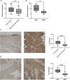
The expression of CENPF in expanded 30 pairs of bladder cancer tissues and adjacent normal tissues and IHC staining of CENPF in patients. (A)(B) CENPF expression is higher in tumor tissues than in adjacent normal tissues, and higher in MIBC than NMIBC(P<0.05). (C) CENPF is not/weak stained in typical urothelium. CENPF is mainly moderate to strong stained in tumor cells(P < 0.05). (D) NMIBC, weak(rarely) to moderate(almost) stained; MIBC, moderate to strong stained. Increased staining of CENPF was detected of tumor cells in MIBC compared with NMIBC(P < 0.05).
Fig. 3
Fig. 3
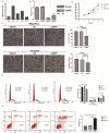
The effects of reduced CENPF expression on bladder cancer cells. (A) The expression levels of CENPF in different bladder cell lines (T24,5637, J82, EJ and T921 cells) were measured by qRT-PCR. (B) The siRNA1 was better transfection efficiency through qRT-PCR and the Western Blot experiment. (C) The proliferation capacity of T24 cells was measured by CCK-8 assays after interference with MOCK, NC and siRNA1 at 0 d, 1 d, 2d,3d,4d and 5d(P<0.05). (D) The cell migration and invasion of T24 cells were evaluated by transwell assay at 100x after 24 h. Numbers of migration and invasive cells compared to mock, NC groups were measured in the right charts. Data were presented as means ± SD; n = 3. SD: standard deviation. (E) Cell cycle assays of T24 cells. The proportions of cells in the G1, G2, and S phase for the three groups were shown in the right charts. (F) Flow cytometry analysis of apoptosis of cells. The apoptosis rates of T24 cells in the three groups were shown in the right charts (P < 0.05).
Fig. 4
Fig. 4
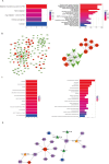
Functional enrichment analyses, protein-protein interaction networks, and lncRNA-microRNA-mRNA (ceRNAs) interactions. (A) The KEGG pathway analyses for both upregulated and downregulated genes are shown. The left side displays the pathway enrichment outcomes for upregulated mRNAs, while the right side presents the pathway enrichment results for downregulated mRNAs. The x-axis denotes the quantity of enriched genes, and the y-axis identifies the KEGG pathway names. The p-value indicates the significance threshold, with the color gradient transitioning from blue to red signifying a reduction in p-values and an increase in significance. (B) The protein-protein interaction (PPI) network and module analysis is depicted. The left side illustrates the PPI network established from 129 overlapping target genes, whereas the right side focuses on the PPI network involving the target gene CENPF and TMEM132A. The green downward triangles signify downregulated mRNAs, while the red circles denote upregulated mRNAs. The size of the nodes correlates with the degree value magnitude. (C) Enrichment analysis of CENPF and TMEM132A gene cluster. The left side reveals the pathway enrichment results, and the right side showcases the Biological Process (BP) enrichment outcomes. The x-axis represents the number of enriched genes, while the y-axis specifies the names of the pathways or biological processes. The p-value serves as the significance threshold, with colors ranging from blue to red indicating decreasing p-values and increasing significance. (D) The lncRNA-miRNA-mRNA (ceRNAs) network for the CENPF gene is illustrated. Red circles indicate upregulated genes, orange upward triangles represent upregulated miRNAs, green upward triangles denote downregulated miRNAs, blue squares signify downregulated lncRNAs, and purple squares represent upregulated lncRNAs.

Similar articles

References

    1. Sanguedolce, F. et al. HER2 expression in bladder cancer: a focused view on its diagnostic, prognostic, and predictive role. Int. J. Mol. Sci.24, 145. 10.3390/ijms24043720 (2023). - PMC - PubMed
    1. Li, J. et al. Construction of a novel mRNA-signature prediction model for prognosis of bladder cancer based on a statistical analysis. BMC Cancer. 21, 858. 10.1186/s12885-021-08611-z (2021). - PMC - PubMed
    1. Contieri, R. et al. Accuracy of the European association of urology (EAU) NMIBC 2021 scoring model in predicting progression in a large cohort of HG T1 NMIBC patients treated with BCG. Minerva Urol. Nephrol.75, 180–187. 10.23736/s2724-6051.22.04953-9 (2023). - PubMed
    1. Babjuk, M. et al. European association of urology guidelines on Non-muscle-invasive bladder Cancer (Ta, T1, and carcinoma in Situ). Eur. Urol.81, 75–94. 10.1016/j.eururo.2021.08.010 (2022). - PubMed
    1. Cathomas, R. et al. The 2021 updated European association of urology guidelines on metastatic urothelial carcinoma. Eur. Urol.81, 95–103. 10.1016/j.eururo.2021.09.026 (2022). - PubMed

MeSH terms

LinkOut - more resources