

Role of Neo-Sinus on Thrombogenicity of Aortic Valve Prostheses: Experimental Proof-of-Concept Study
- PMID: 40660009
- PMCID: PMC12528357
- DOI: 10.1007/s13239-025-00792-z
Role of Neo-Sinus on Thrombogenicity of Aortic Valve Prostheses: Experimental Proof-of-Concept Study
Abstract
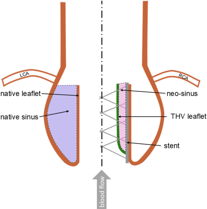
Purpose: Transcatheter aortic valve replacement (TAVR) is the standard treatment for patients with aortic diseases at high surgical risk. Transcatheter heart valve prostheses (THV) are inserted into the aortic valve, creating a new area between the native and artificial leaflets. This area, known as neo-sinus, increases the thrombogenicity of THVs. But there is a lack of testing methods that evaluate thrombogenicity in vitro.
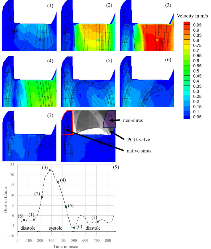
Methods: To analyze the flow field within the native sinus and the neo-sinus, Particle Image Velocimetry (PIV) was performed with a thrombosis tester. Additionally, a comparative study was conducted with porcine blood on two polycarbonate urethane valves, with and without neo-sinus, respectively. Blood samples collected every hour were analyzed for platelet count, coagulation via ROTEM parameters, and plasma-free hemoglobin. Thrombus formation was detected optically.
Results: The PIV measurements yield a physiological flow field in the aortic root that were consistent with those reported in literature. The analyzed blood parameters reveal no obvious difference between the valve with neo-sinus and the valve without. A higher amount of thrombus material for the valve with neo-sinus was found.
Conclusion: The visualized flow field shows low velocities and stagnation zones due to the presence of native leaflets. Clot formation at the heart valve prostheses are in accordance with in-vivo findings. The benchmark of the two valves indicates an increased thrombogenic potential due to the neo-sinus. The thrombosis tester simulates the natural environment after TAVR. Thereby, newly developed THVs can be evaluated in vitro and consequently optimized regarding their thrombogenicity.
Keywords: Blood test; In-vitro testing; Neo-sinus; PIV measurements; Thrombogenicity; Transcatheter aortic valve replacement.
© 2025. The Author(s).
Conflict of interest statement
Declarations. Ethics Approval, Consent to Participate & to Publish: Not applicable. Conflict of Interest: The authors have no competing interests to declare that are relevant to the content of this article.
Figures


References
-
- Popma JJ, Deeb GM, Yakubov SJ, Mumtaz M, Gada H, O’Hair D, et al. Transcatheter Aortic-Valve Replacement with a Self-Expanding Valve in Low-Risk Patients. N Engl J Med. 2019;380:1706–15. 10.1056/NEJMoa1816885. - PubMed
-
- Siontis GCM, Overtchouk P, Cahill TJ, Modine T, Prendergast B, Praz F, et al. Transcatheter aortic valve implantation vs. surgical aortic valve replacement for treatment of symptomatic severe aortic stenosis: an updated meta-analysis. Eur Heart J. 2019;40:3143–53. 10.1093/eurheartj/ehz275. - PubMed
-
- Mack MJ, Leon MB, Thourani VH, Makkar R, Kodali SK, Russo M, et al. Transcatheter Aortic-Valve Replacement with a Balloon-Expandable Valve in Low-Risk Patients. N Engl J Med. 2019;380:1695–705. 10.1056/NEJMoa1814052. - PubMed
-
- Vahanian A, Beyersdorf F, Praz F, Milojevic M, Baldus S, Bauersachs J, et al. 2021 ESC/EACTS Guidelines for the management of valvular heart disease. Eur Heart J. 2022;43:561–632. 10.1093/eurheartj/ehab395. - PubMed
-
- Chakravarty T, Søndergaard L, Friedman J, Backer O de, Berman D, Kofoed KF, et al. Subclinical leaflet thrombosis in surgical and transcatheter bioprosthetic aortic valves: an observational study. Lancet (London, England). 2017;389:2383–92. 10.1016/S0140-6736(17)30757-2. - PubMed
MeSH terms
LinkOut - more resources
Full Text Sources
Medical
