

Simultaneous involvement of pigmented villonodular synovitis in the left shoulder glenohumeral joint and ankle joint: a rare case report
- PMID: 40660237
- PMCID: PMC12257791
- DOI: 10.1186/s12891-025-08936-x
Simultaneous involvement of pigmented villonodular synovitis in the left shoulder glenohumeral joint and ankle joint: a rare case report
Abstract
Background: Pigmented Villonodular Synovitis, although uncommon, can lead to significant joint destruction if not diagnosed and treated early. Concurrent polyarticular presentation in adults is exceedingly rare, appearing in less than 1% of cases.
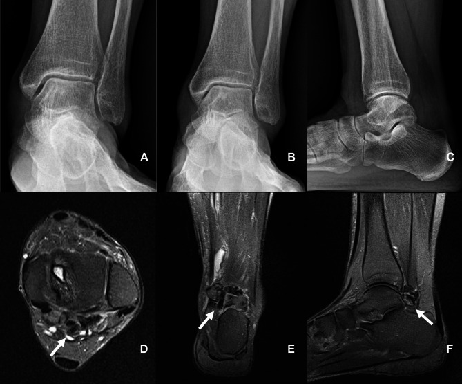
Case presentation: This case report presents a unique and rare occurrence of Pigmented Villonodular Synovitis involving both the ankle and shoulder joints simultaneously in an adult patient. The patient initially developed a diffuse form Pigmented Villonodular Synovitis in the left ankle, which was managed with total synovectomy at another tertiary hospital. Two months later, a separate localized form Pigmented Villonodular Synovitis was diagnosed in the left glenohumeral joint, necessitating arthroscopic marginal resection at our hospital. At the one-year follow-up, the magnetic resonance imaging of the left shoulder showed no signs of Pigmented Villonodular Synovitis recurrence, with full range of motion and no pain. However, the magnetic resonance imaging of the left ankle revealed a recurrence of the Pigmented Villonodular Synovitis, despite the range of motion remaining within normal limits.
Conclusions: This case demonstrates the importance of considering multiarticular Pigmented Villonodular Synovitis when symptoms manifest in multiple joints. Early diagnosis and appropriate intervention are critical to prevent joint destruction and optimize patient outcomes.
Keywords: Case report; Marginal resection; Multiarticular pigmented villonodular synovitis; Pigmented villonodular synovitis; Shoulder; Synovectomy.
© 2025. The Author(s).
Conflict of interest statement
Declarations. Ethics approval and consent to participate: The patient agreed to participate in this study. Informed consent was obtained from all individual participants included in the study. All procedures were conducted according to the Declaration of Helsinki. Consent for publication: Written informed consent was obtained from the patient to publish her case-related information and images. Competing interests: The authors declare no competing interests.
Figures


Similar articles
-
What Are the Recurrence Rates, Complications, and Functional Outcomes After Multiportal Arthroscopic Synovectomy for Patients With Knee Diffuse-type Tenosynovial Giant-cell Tumors?Clin Orthop Relat Res. 2024 Jul 1;482(7):1218-1229. doi: 10.1097/CORR.0000000000002934. Epub 2023 Dec 28. Clin Orthop Relat Res. 2024. PMID: 38153106 Free PMC article.
-
Giant cell tumor of tendon sheath: Open surgery or arthroscopic synovectomy? A systematic review of the literature.Orthop Traumatol Surg Res. 2017 Sep;103(5):809-814. doi: 10.1016/j.otsr.2017.03.016. Epub 2017 Apr 17. Orthop Traumatol Surg Res. 2017. PMID: 28428036
-
Satisfactory functional outcomes and low recurrence rates at a mean 10-year follow-up after combined staged synovectomy and external radiotherapy for diffuse pigmented villonodular synovitis of the knee.J ISAKOS. 2025 Aug;13:100907. doi: 10.1016/j.jisako.2025.100907. Epub 2025 May 24. J ISAKOS. 2025. PMID: 40419142
-
[Arthroscopic treatment of pigmented villonodular synovitis of the ankle: a clinical case report and review].Medwave. 2019 May 27;19(4):e7641. doi: 10.5867/medwave.2019.04.7641. Medwave. 2019. PMID: 31150371 Review. Spanish.
-
Pigmented villonodular synovitis of the shoulder.Orthopedics. 1989 May;12(5):715-8. doi: 10.3928/0147-7447-19890501-10. Orthopedics. 1989. PMID: 2657682 Review.
References
-
- Doherty BT, Morrison WB. Other Intra-articular synovial pathology. Musculoskelet Imaging. 2024:1–6.
-
- Chassaignac M. Cancer de la gaine des tendons. Gazette des Hôpitaux Civils et Militaires. 1852.
-
- Jaffe HL. Pigmented villonodular synovitis, bursitis and tenosynovitis. Arch Pathol. 1941;31:731–65.
-
- Flandry F, Hughston J. Pigmented villonodular synovitis. J Bone Joint Surg. 1987;69(6):942–9. - PubMed
-
- Tyler WK, Vidal AF, Williams RJ, Healey JH. Pigmented villonodular synovitis. J Am Acad Orthop Surg. 2006;14(6):376–85. - PubMed
Publication types
MeSH terms
LinkOut - more resources
Full Text Sources
