Review
. 2025 Jul 15;16(1):368.
doi: 10.1186/s13287-025-04511-2.

Mesenchymal stem cell-derived exosomes as a potential therapeutic strategy for ferroptosis

Affiliations
Review

Mesenchymal stem cell-derived exosomes as a potential therapeutic strategy for ferroptosis

Mohammed Zayed et al. Stem Cell Res Ther. .

Abstract

Ferroptosis, a regulated type of cell death directed by iron-dependent lipid peroxidation, is associated with a variety of pathological diseases. Recent findings have highlighted the therapeutic potential of mesenchymal stem cell-derived exosomes (MSC-Exos) in modulating ferroptosis. These nano-sized extracellular vesicles carry bioactive substances, including proteins, lipids, and microRNAs, which regulate vital pathways related to ferroptosis, such as reactive oxygen species production, glutathione metabolism, and lipid peroxidation. Preclinical studies suggest that MSC-Exos can alleviate ferroptosis-induced damage by enhancing antioxidant defenses, mitigating oxidative stress, upregulating anti-ferroptotic regulators, and suppressing lipid peroxidation. Notably, in cancer, MSC-Exos may protect non-malignant tissues from chemotherapy-induced ferroptosis. By exploiting their regenerative and immunomodulatory properties, MSC-Exos offer a promising therapeutic platform for targeting ferroptosis in diverse pathological conditions. This review summarizes the biological and functional characteristics of MSC-Exos, elucidates their roles in ferroptosis regulation across multiple disease models, and discusses current challenges and future directions for clinical translation.

Keywords: Cancer; Cell death; Exosomes; Ferroptosis; Mesenchymal stem cells; Neurodegenerative diseases; Oxidative stress; Regenerative medicine.


Conflict of interest statement

Declarations. Ethics approval and consent to participate: Not applicable. Consent for publication: Not applicable. Competing interests: The authors declare that they have no competing interests.

Figures

Fig. 1
Fig. 1
Schematic representation of the biogenesis of exosomes released from mesenchymal stem cells (MSC-Exos). (A) The biogenesis of Exos begins with the creation of early Exos through the invagination of the plasma membrane, followed by the development of late Exos through the selection of specific cargo. It ultimately leads to the formation of multivesicular bodies (MVBs). MVBs contain intraluminal vesicles (ILVs). (B) The molecular components of endosomal sorting complexes required for transport (ESCRT-0, -I, -II, -III) and their interaction to form and release ILVs have been illustrated. Created with BioRender.com
Fig. 2
Fig. 2
The composition and therapeutic roles of mesenchymal stem cell-derived exosomes (MSC-Exos). (A) MSC-Exos contain cytokines, proteins, lipids, mRNAs, miRNAs, and ncRNAs. MSC-Exos possesses several therapeutic benefits, such as immunomodulatory effects, promotion of cell survival, differentiation and migration, anti-apoptotic activities, and the ability to enhance angiogenesis. (B) The uptake of Exos by the recipient cell through direct interaction with specific receptors on the plasma membrane, plasma membrane fusion, and by endocytosis. After uptake, Exos deliver exosomal cargo to specific organelles, such as the endoplasmic reticulum (ER) (1); Exos cargo release by fusion with the endosomal membrane (2); undergo lysosomal degradation (3); fuse with a lysosome and releasing the soluble cargo of Exos (4); be trafficked back to the plasma membrane for re-secretion (5). Created with BioRender.com
Fig. 3
Fig. 3
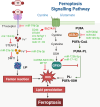
The ferroptosis pathways. The primary metabolic processes involved in ferroptosis can be classified into three main categories: iron metabolism, the GSH/GPX4 pathway, and lipid peroxidation. Ferrous ions (Fe2+) contribute to the accumulation of lipid peroxides via the Fenton reaction and the oxidation of lipids. Cystine is reduced to cysteine, which is utilized to synthesize glutathione (GSH). GPX4 can reduce harmful lipid peroxides. Long-chain fatty acid-CoA ligase 4 (ACSL4) facilitates the attachment of polyunsaturated fatty acids (PUFAs) to phospholipids, resulting in the formation of polyunsaturated fatty acid-containing phospholipids (PUFA-PLs), which can subsequently be oxidized to lipid peroxides. Different cargo molecules of mesenchymal stem cell-derived exosomes can target different ferroptosis-related signaling pathways. Created with BioRender.com

References

    1. Dixon SJKM, Lemberg MR, Lamprecht, et al. Ferroptosis: an iron-dependent form of nonapoptotic cell death. Cell. 2012;149(5):1060–72. 10.1016/j.cell.2012.03.042. - PMC - PubMed
    1. Stockwell BRJP, Friedmann Angeli H, Bayir, et al. Ferroptosis: A regulated cell death Nexus linking metabolism, redox biology, and disease. Cell. 2017;171(2):273–85. 10.1016/j.cell.2017.09.021. - PMC - PubMed
    1. Yan JL, Bao H, Liang, et al. A druglike Ferrostatin-1 analogue as a ferroptosis inhibitor and photoluminescent Indicator. Angew Chem Int Ed. 2025. 10.1002/anie.202502195. e202502195.DOI. - PubMed
    1. Zhang JF, Lin Y, Xiao, et al. Discovery and optimization of 1,2,4-triazole derivatives as novel ferroptosis inhibitors. Eur J Med Chem. 2025;284:117192. 10.1016/j.ejmech.2024.117192. - PubMed
    1. Alves FD, Lane TPM, Nguyen AI, Bush S, Ayton. In defence of ferroptosis. Signal Transduct Target Therapy. 2025;10(1). 10.1038/s41392-024-02088-5. p. 2. - PMC - PubMed

Substances

LinkOut - more resources