Review
. 2025 Jul 15;16(1):366.
doi: 10.1186/s13287-025-04441-z.

Diabetes and periodontitis: the role of a high-glucose microenvironment in periodontal tissue cells and corresponding therapeutic strategies

Affiliations
Review

Diabetes and periodontitis: the role of a high-glucose microenvironment in periodontal tissue cells and corresponding therapeutic strategies

Dan-Dan Xiang et al. Stem Cell Res Ther. .

Abstract

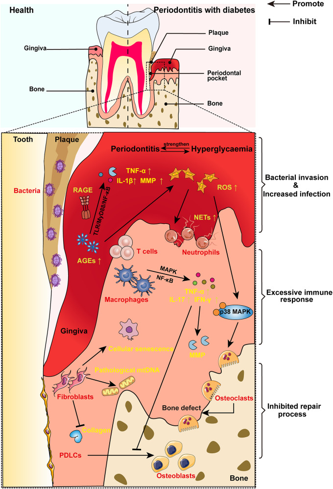
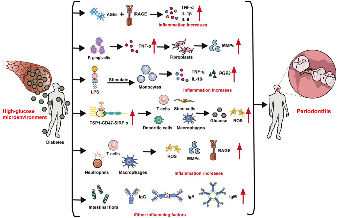
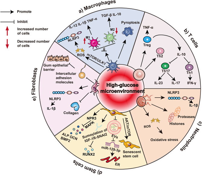
Background: Periodontitis is a chronic inflammatory disease that leads to the destruction of periodontal tissues and diabetes is a metabolic disease characterized by hyperglycemia. Both of these diseases affect many people worldwide. Epidemiological data have revealed a close relationship between periodontitis and diabetes. In particular, the high-glucose microenvironment plays an important role in the relationship between these two chronic inflammatory diseases, which makes it difficult to mitigate the progression of periodontitis and restore periodontal tissue in diabetic patients. Anti-inflammatory and regenerative processes are of fundamental importance for periodontal treatment and are mediated by diverse cell populations, including macrophages, T cells, neutrophils, stem cells, and fibroblasts that reside within periodontal tissues.

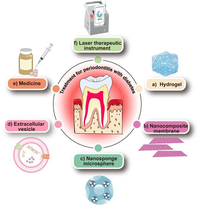
Main body: This review summarizes the interaction between diabetes mellitus and periodontitis, and illustrates that the high-glucose microenvironment aggravates periodontal homeostasis. Furthermore, the mechanism by which the high-glucose microenvironment regulates the involvement of various cells in the destruction of periodontal tissue, leading to the significant inhibition of tissue regeneration and recovery, is discussed. On this basis, the current therapeutic strategies that can be used to target cells are summarized to improve the regeneration and repair processes in the high-glucose microenvironment.

Conclusion: In this review, we assess how metabolic dysregulation mediated by a high-glucose microenvironment exacerbates inflammatory damage and inhibits tissue repair. A deeper understanding of the effects of a high-glucose microenvironment on periodontal tissue cells is essential for developing new therapeutic strategies to restore the structure and function of periodontal tissue.

Keywords: High-glucose microenvironment; Immune cells; Periodontitis; Stem cells; Therapeutic strategies.


Conflict of interest statement

Declarations. Ethics approval and consent to participate: Not applicable. Consent for publication: Not applicable. Competing interests: The authors declare that they have no competing interests.

Figures

Fig. 1
Fig. 1
Influence of a high-glucose microenvironment on periodontal conditions. High glucose levels drive the pathological accumulation of advanced glycosylation end products (AGEs) and reactive oxygen species (ROS), intensifying the inflammatory response triggered by bacteria. This self-amplifying inflammatory cascade induces an excessive immune response, such as activation of the TLR/MyD88/NF-κB signaling pathway when AGEs bind to the receptor of AGEs (RAGE), upregulating proinflammatory cytokines such as tumor necrosis factor-α (TNF-α), interleukin (IL)-1β, and matrix metalloproteinases (MMPs). AGEs can also promote the generation of ROS. ROS trigger neutrophils to form excessive neutrophil extracellular traps (NETs) formation, which hinders the resolution of inflammation. Additionally, ROS can activate the p38 MAPK signaling pathway and then mediate osteoclastogenesis, further exacerbating alveolar bone loss. Concurrently, the glucose-dysregulated microenvironment polarizes T cells and macrophages toward proinflammatory phenotypes, characterized by excessive secretion of IL-17, interferon-γ (IFN-γ), and TNF-α. These inflammatory mediators promote the production of MMP, degrade the extracellular matrix, and inhibit the differentiation of osteoblasts to limit the repair process. In addition, abnormal glucose levels impair the normal functions of fibroblasts, promoting their aging, inducing the production of pathological mtDNA, and impairing collagen synthesis. These linked mechanisms form a loop between high-glucose stimuli and periodontal destruction, with therapeutic targets identified at checkpoints
Fig. 2
Fig. 2
Mechanisms underlying diabetes-induced progression of periodontitis. The influence of high-glucose microenvironments triggers the activation of immune cells, accelerating the development of periodontitis. The interaction of AGEs with RAGE leads to increased inflammation and pro-oxidative stress by increasing the secretion of proinflammatory factors (e.g., TNF-α, IL-1β and IL-6). The exacerbated inflammatory cascade elicited by Porphyromonas gingivalis (P. gingivalis) contributes further to the release of TNF-α, prompting fibroblasts to generate enzymes that degrade MMPs. The high-glucose microenvironment enhances the LPS-stimulated proinflammatory effects on monocytes by triggering an increase in the secretion of proinflammatory cytokines (such as TNF-α, IL-1β and prostaglandin E2 (PGE2)). The TSP1-CD47-SIRPα axis regulates the functions of immune cells and stem cells, etc., resulting in the dysregulation of glucose homeostasis and the increase of ROS. A high-glucose microenvironment decreases the activity and chemotaxis of polymorphonuclear neutrophils (PMNs) but significantly increases their secretion of MMPs and ROS, exacerbating periodontitis-induced tissue destruction. Moreover, this process involves macrophage apoptosis, leading to increased production of RAGE. In addition, other influencing factors include the intestinal flora and immune proteins such as IgG, IgA and IgM, which also play roles in the interaction between the high-glucose microenvironment and periodontitis. The irreversible pathological feed-forward loop drives progressive alveolar bone resorption and collagen matrix degradation in diabetic individuals
Fig. 3
Fig. 3
The high-glucose microenvironment induces intricate complex cellular responses and signaling pathways. (a) When stimulated by a high-glucose microenvironment, macrophages tend to differentiate into M1 macrophages, with an increase in the number of M1 macrophages and a decrease in the number of M2 macrophages. M1 macrophage activation elicits inflammatory processes and the release of cytokines such as IL-12, IL-1β, and TNF-α; conversely, M2 macrophages secrete TGF-β and IL-10. The high-glucose microenvironment can lead to macrophage pyroptosis. The mammalian target of rapamycin (mTOR)/UNC-52-like kinase 1 (ULK1) pathway is also activated, thereby impairing macrophage autophagy and leading to increased ROS secretion and the activation of NOD-like receptor protein-3 (NLRP3) inflammatory vesicles. (b) Regulatory T cells (Tregs) secrete TNF-α and IL-10, whereas Th2 cells primarily release IL-10. IL-10 acts as a regulatory cytokine that inhibits Th1 and Th17 responses. Th17 cells produce IL-23 and IL-17, and Th1 cells secrete IFN-γ, contributing to specific immune responses. (c) Neutrophil activation via the NLRP3 inflammasome enhances inflammation via the release of proteases and histones, exacerbating oxidative stress through ROS generation. (d) In mesenchymal stem cells (MSCs), high glucose reduces the secretion of exosomes and the release of miR-129-3p, ultimately leading to endoplasmic reticulum (ER) dysfunction. High glucose also negatively affects MSCs, leading to cellular senescence through the AKT/mTOR signaling pathway. The high-glucose microenvironment upregulated the natriuretic peptide receptor 3 (NPR3)-mediated mitogen-activated protein kinase (MAPK) pathway while affecting the expression levels of key factors such as alkaline phosphatase (ALP), osteocalcin (OCN) and bone morphogenetic protein 2 (BMP2). Sumoylation of IGF-1R-SNAI2 can reduce the level of RUNX2. (e) Furthermore, impaired expression of fibroblast adhesion molecules disrupts the integrity of the gingival epithelial barrier. This disruption is accompanied by the activation of inflammatory mediators, reduced collagen synthesis, and constraints on fibroblast proliferation and migration processes
Fig. 4
Fig. 4
Multifaceted strategies incorporating advanced biomedical interventions for diabetes-related periodontitis. (a) Hydrogel: Hydrogel can be utilized as a foundation platform to fabricate innovative biomaterials tailored for periodontal tissue regeneration. This includes the formulation of heat-sensitive or glucose-responsive hydrogels designed to enhance the cellular response of immune cells or stem cells. (b) Nanocomposite membrane: The unique characteristics of nanomaterials within nanocomposite membranes are harnessed to modulate the electrical microenvironment, fostering the polarization of macrophages toward the reparative M2 phenotype. (c) Microsphere: Drug-loaded microspheres can be uced to ameliorate the cellular dysfunction induced by the high-glucose microenvironment. For example, microspheres containing therapeutic agents such as Ex-4 can be utilized to enhance cellular functions. (d) Extracellular vesicles: Explore the therapeutic potential of extracellular vesicles in orchestrating the physiological responses of target cells. Engineering extracellular vesicles offers a promising avenue to mitigate the detrimental inflammatory cascades triggered by elevated glucose levels. (e) Medicine: Integrate pharmaceutical agents into conventional periodontal treatment protocols or develop bioactive materials to enhance therapeutic outcomes. (f) Oral laser treatment: Laser-based interventions are implemented to facilitate angiogenesis and collagen synthesis, thereby fostering the regeneration and restoration of periodontal tissues through the promotion of blood vessel growth and collagen fiber synthesis

Similar articles

References

    1. GBD 2021 Diabetes Collaborators. Global, regional, and National burden of diabetes from 1990 to 2021, with projections of prevalence to 2050: a systematic analysis for the global burden of disease study 2021. Lancet. 2023;402(10397):203–34. - PMC - PubMed
    1. GBD 2017 Disease and Injury Incidence and Prevalence Collaborators. Global, regional, and National incidence, prevalence, and years lived with disability for 354 diseases and injuries for 195 countries and territories, 1990–2017: a systematic analysis for the global burden of disease study 2017. Lancet. 2018;392(10159):1789–858. - PMC - PubMed
    1. Luo LS, Luan HH, Wu L, et al. Secular trends in severe periodontitis incidence, prevalence and disability-adjusted life years in five Asian countries: A comparative study from 1990 to 2017. J Clin Periodontol. 2021;48(5):627–37. - PubMed
    1. Preshaw PM, Alba AL, Herrera D, et al. Periodontitis and diabetes: a two-way relationship. Diabetologia. 2012;55(1):21–31. - PMC - PubMed
    1. Lalla E, Papapanou PN. Diabetes mellitus and periodontitis: a Tale of two common interrelated diseases. Nat Rev Endocrinol. 2011;7(12):738–48. - PubMed

LinkOut - more resources