

TLR3-overexpressing umbilical cord mesenchymal stromal cells suppress immune responses to attenuate high-risk corneal transplantation rejection
- PMID: 40660345
- PMCID: PMC12261691
- DOI: 10.1186/s13287-025-04510-3
TLR3-overexpressing umbilical cord mesenchymal stromal cells suppress immune responses to attenuate high-risk corneal transplantation rejection
Abstract
Background: The success rates of high-risk corneal transplantation are significantly hindered by immunological rejection. Umbilical cord mesenchymal stem cells (UC-MSCs) have emerged as a potential solution to improve graft outcomes. Toll-like receptors (TLRs) play pivotal roles in the immune response, however, their specific functions in UC-MSC-based treatments for corneal graft rejection requires further investigation.
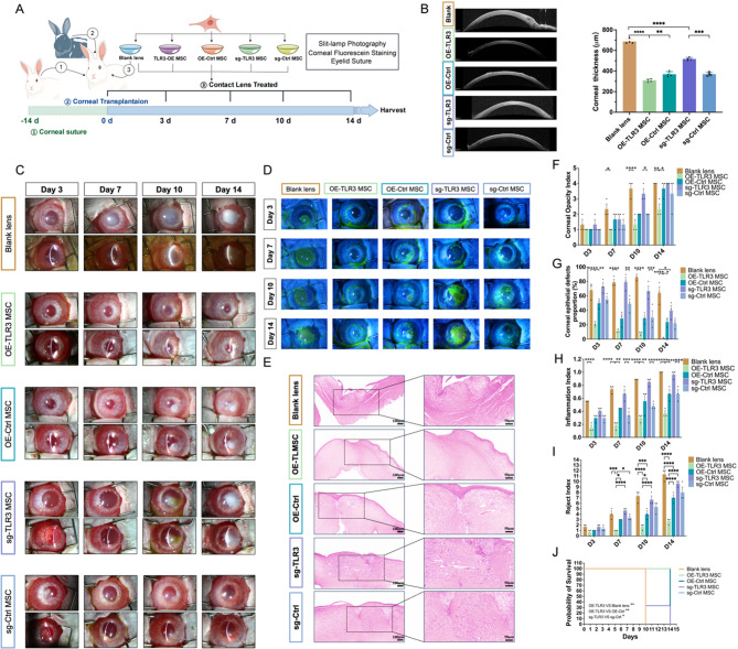
Methods: New Zealand rabbits that underwent high-risk corneal transplantation were treated with UC-MSC-coated contact lenses (MSCohi-O), while blank lenses and untreated grafts served as controls. Clinical manifestations related to rejection were assessed. RNA-seq was performed on the grafts. GSEA, CIBERSORT, and flow cytometry analyses were performed to investigate the activation of signaling pathways and immune changes. The microarray dataset GSE68610, which contains transcriptome data for normal human UC-MSCs and UC-MSCs treated with cytokines, was analyzed to evaluate TLR expression and identify the key factors involved in the anti-inflammatory effect of UC-MSCs. Overexpression and knockout of TLR3 were performed in UC-MSCs, and the therapeutic effects of TLR3-activated and -inactivated UC-MSCs were compared in a high-risk corneal transplantation model.
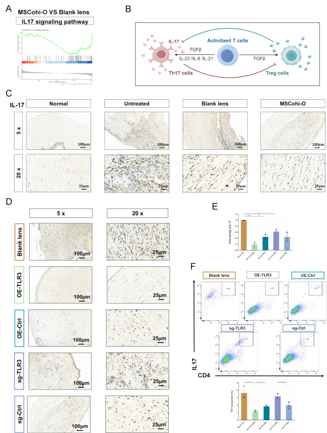
Results: MSCohi-O treatment alleviated corneal opacity and edema, inhibited neovascularization, promoted epithelialization, and prolonged the survival time of corneal grafts (all p < 0.05). GSEA revealed that the allograft rejection pathway was upregulated in untreated grafts and downregulated in UC-MSC-treated grafts. Increased numbers of Tregs and decreased numbers of Th17 cells were observed in UC-MSC-treated corneas. An analysis of the GSE68610 dataset revealed that TLR3 expression was upregulated in cytokine-activated UC-MSCs, suggesting that TLR3 is a potential regulator of the immunosuppression function of UC-MSCs. TLR3-overexpressing UC-MSCs exhibited enhanced suppression of rejection in high-risk corneal transplants, whereas TLR3 knockdown diminished these effects.
Conclusions: This study shows that the localized application of UC-MSC-coated contact lenses is capable of inhibiting rejection in high-risk corneal transplantation. TLR3-overexpressing UC-MSCs have enhanced antirejection and immunomodulatory effects. This research could offer a safer and more effective therapeutic strategy to prevent corneal transplant rejection.
Keywords: Corneal transplantation rejection; Immune; Mesenchymal stromal cells; TLR3.
© 2025. The Author(s).
Conflict of interest statement
Declarations. Ethics approval and consent to participate: All animal experiments for this study were in accordance with the Animal Research: Reporting of In Vivo Experiments (ARRIVE) guidelines 2.0. All procedures were approved by the Ethics Committee of Zhongshan Ophthalmic Center Animal Care and Ethics Committee. The research project titled “Study on the therapeutic effect and mechanism of stem cells and NU7441 in ocular surface and fundus diseases” was approved on November 01, 2023 (ethical approval number: O2023048). Human UC-MSCs related procedures were approved by the Ethics Committee of Third Affiliated Hospital, Sun Yat-sen University. The research project titled “Research on the Role and Mechanism of Mesenchymal Stem Cell Therapy in Treating Corneal Transplant Rejection and Dry Eye Syndrome” was approved on February 07, 2024 (ethical approval number: A2024-78-01). The patients provided written informed consent for the use of samples. Consent for publication: Not applicable. Competing interests: The authors declare no competing interests. Artificial intelligence: The authors declare that they have not used Artificial Intelligence in this study.
Figures


Similar articles
-
Mesenchymal stem cell secretome alters gene expression and upregulates motility of human endometrial stromal cells.Reproduction. 2023 Jul 5;166(2):161-174. doi: 10.1530/REP-22-0485. Print 2023 Aug 1. Reproduction. 2023. PMID: 37252830 Free PMC article.
-
Interleukin-1 receptor antagonist overexpression in mesenchymal stem cells improves hemorrhagic cystitis outcomes via HtrA serine peptidase 3.Stem Cell Res Ther. 2025 Jul 1;16(1):337. doi: 10.1186/s13287-025-04443-x. Stem Cell Res Ther. 2025. PMID: 40598340 Free PMC article.
-
Cell Sheets Formation Enhances Therapeutic Effects of Human Umbilical Cord Mesenchymal Stem Cells on Spinal Cord Injury.CNS Neurosci Ther. 2024 Dec;30(12):e70163. doi: 10.1111/cns.70163. CNS Neurosci Ther. 2024. PMID: 39670537 Free PMC article.
-
Sex and gender as predictors for allograft and patient-relevant outcomes after kidney transplantation.Cochrane Database Syst Rev. 2024 Dec 19;12(12):CD014966. doi: 10.1002/14651858.CD014966.pub2. Cochrane Database Syst Rev. 2024. PMID: 39698949
-
Systematic review and meta-analysis of mesenchymal stromal/stem cells as strategical means for the treatment of COVID-19.Ther Adv Respir Dis. 2023 Jan-Dec;17:17534666231158276. doi: 10.1177/17534666231158276. Ther Adv Respir Dis. 2023. PMID: 37128999 Free PMC article.
References
-
- Figueiredo GS, Jones MN, Krishna Y, Figueiredo FC, Larkin DF, Kaye SB, National Health Service, B., and, Advisory TOT. G. (2015). Transplant rejection following endothelial keratoplasty and penetrating keratoplasty in the United Kingdom: incidence and survival. Am J Ophthalmol 160, 416–421. 10.1016/j.ajo.2015.06.012 - PubMed
-
- Vanathi M, Raj N, Kusumesh R, Aron N, Gupta N, Tandon R. Update on pediatric corneal diseases and keratoplasty. Surv Ophthalmol. 2022;67:1647–84. 10.1016/j.survophthal.2022.07.010. - PubMed
MeSH terms
Substances
Grants and funding
- 2021GH08/Guangzhou Huangpu International Science and Technology Collaboration Project
- 2024A03J0278/Guangzhou Science and Technology Planning Project
- 2023A1515110232/Basic and Applied Basic Research Foundation of Guangdong Province
- 2023/Open Research Funds of the State Key Laboratory of Ophthalmology
LinkOut - more resources
Full Text Sources
