Review
. 2025 Jul 15;13(1):97.
doi: 10.1186/s40364-025-00807-w.

T cells in cancer: mechanistic insights and therapeutic advances

Affiliations
Review

T cells in cancer: mechanistic insights and therapeutic advances

Jingjing Pu et al. Biomark Res. .

Abstract

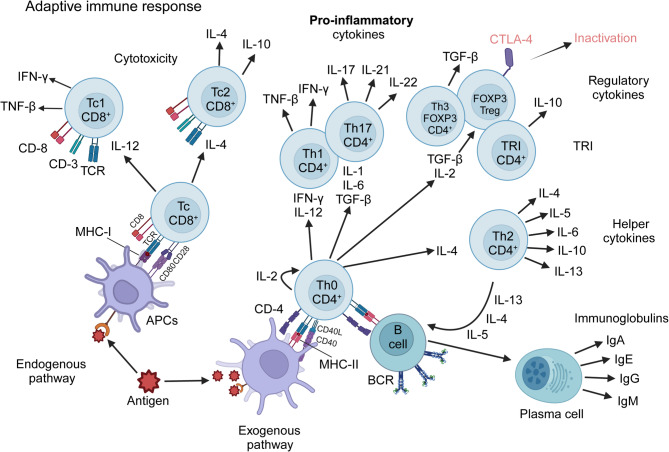
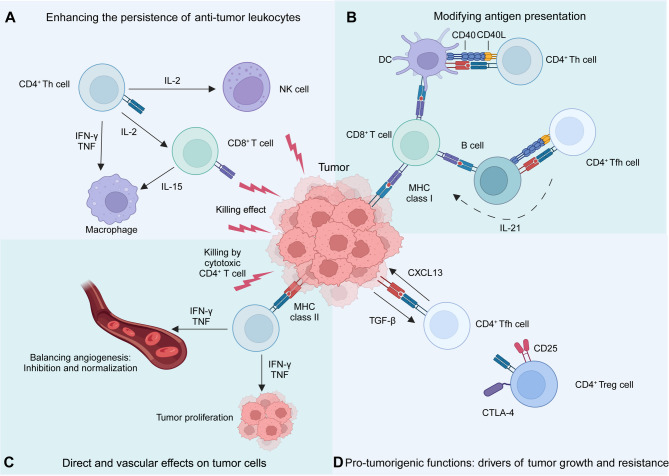
T cells are central players in the fight against cancer, capable of recognizing and destroying tumor cells. However, tumors often find ways to evade this immune response, creating challenges for effective treatment. In this review, we explore how different T cell subsets—including cytotoxic T cells, helper T cells, regulatory T cells, and unconventional T cells—contribute to tumor progression or suppression. We also delve into key mechanisms, such as immune checkpoints and metabolic pathways, that shape T cell behavior in the tumor microenvironment. Advances in cancer immunotherapy, including immune checkpoint inhibitors (ICIs), T cell engagers (TCEs), adoptive T cell therapies (ACTs), chimeric antigen receptor (CAR) T cell therapies, and cancer vaccines, have transformed cancer treatment and provided new hope for patients. However, challenges such as treatment resistance, limited efficacy in solid tumors, and therapy-associated toxicities remain significant barriers to broader clinical success. We discuss innovative strategies to tackle these challenges, including combination therapies and next-generation T cell engineering approaches. By connecting the biology of T cells with cutting-edge therapeutic advances, this review aims to inspire progress in the development of more effective and personalized cancer treatments.

Keywords: Adoptive T cell therapy; Cancer immunotherapy; Precision treatment; T cells; Tumor microenvironment.


Conflict of interest statement

Declarations. Ethics approval and consent to participate: Not applicable. Consent for publication: Not applicable. Competing interests: The authors declare no competing interests.

Figures

Fig. 1
Fig. 1
Overview of T-cell development in the thymus. Committed lymphoid progenitors originate in the bone marrow and migrate to the thymus, where they differentiate into DN thymocytes, lacking TCR, CD4, and CD8. DN thymocytes progress through four stages: early thymic progenitors (ETPs)/DN1 (CD44+CD25), DN2 (CD44+CD25+), DN3 (CD44CD25+), and DN4 (CD44CD25). During the DN2 to DN4 transition, thymocytes express the pre-TCR, consisting of a rearranged TCR β-chain and the invariant pre-Tα chain. Successful pre-TCR signaling drives proliferation and the replacement of the pre-TCR α-chain with a rearranged TCR α-chain, forming a complete αβ-TCR. DP thymocytes interact with MHC-expressing cortical epithelial cells presenting self-peptides. TCR signaling strength determines thymocyte fate: weak signaling causes death by neglect, strong signaling leads to negative selection, and optimal signaling induces positive selection. T cells that bind self-peptide–MHC class I become CD8+ T cells, while those binding MHC class II become CD4+ T cells, maturing for export to peripheral blood and lymphoid tissues. Figure created with BioRender.com
Fig. 2
Fig. 2
Schematic representation of the two major pathways in T cell differentiation. The Th and Tc populations, along with their respective subsets, are activated following antigen uptake and processing by APC, depicted here as a dendritic cell. The processed peptide is presented to the CD8+ population in the context of MHC-I, or to the CD4+ population in the context of MHC-II. This interaction triggers a cascade of lymphoproliferative and differentiative events, influenced by cytokines, which ultimately define the effector functions of these T cells. Figure created with BioRender.com
Fig. 3
Fig. 3
The role of CD8+ cytotoxic T cells in cancer. CTLs play a key role in cancer immunity by eliminating malignant and infected cells. Their activation is influenced by CD4+ T cells and APCs, which promote IL-2 secretion, while IFN-γ drives their maturation. Mature CD8+ T cells generate short-lived effector cells that kill tumor cells through direct cytotoxicity and indirect immune mechanisms. (A) Naïve CD8⁺ T cells recognize antigen presented by professional APCs, supported by CD4⁺ helper T cells, initiating their differentiation into functional cytotoxic T lymphocytes (CTLs). During activation, CD8⁺ T cells upregulate both stimulatory receptors (e.g., CD28, 4-1BB, OX40, ICOS) that promote proliferation and effector functions, and inhibitory receptors (e.g., PD-1, CTLA-4, LAG-3, TIM-3, TIGIT) that restrain excessive activation. Notably, PD-1 expression early after activation helps maintain immune homeostasis. The dynamic balance between these receptors determines the strength, duration, and outcome of the CD8⁺ T cell response, influencing effector activity, exhaustion, and memory development. (B) CTLs eliminate tumor cells through a direct cytotoxic mechanism involving perforin and granzyme. This process relies on cell-to-cell contact, which triggers the release of cytolytic enzymes, including granzyme B. Perforin forms pores in the target cell membrane, allowing granzyme B to enter and initiate apoptotic cell death. (C) Direct tumor cell killing occurs through the interaction between Fas ligand (Fas-L), expressed on CTLs, and its receptor Fas, present on cancer cells. This Fas/Fas-L binding triggers apoptosis in cancer cells via a caspase-dependent pathway. (D) Indirect CD8+ T cell-mediated killing: CTLs can induce indirect, or “bystander,” tumor cell death by secreting cytokines that exert their effects at a distance. For example, TNF-α secretion can activate apoptotic signaling in tumor cells expressing TNF receptors, contributing to immune-mediated tumor clearance. (E) Beyond antigen recognition, co-stimulation, and cytokines, metabolic reprogramming serves as a crucial fourth signal that controls CD8⁺ T cell function. Shifts in pathways like glycolysis, oxidative phosphorylation, and fatty acid metabolism support T cell proliferation, effector activity, and memory formation. Metabolic regulators such as mTOR and AMPK integrate these signals to shape T cell fate and function. Figure created with BioRender.com
Fig. 4
Fig. 4
The role of CD4+ T cells in cancer. CD4+ T cell plasticity shapes innate and adaptive immunity within the TME, secondary lymphoid tissues, and tertiary lymphoid structures. (A) CD4+ T cells support the persistence and function of anti-tumor leukocytes, including NK cells, CD8+ T cells, and myeloid cell populations, through the secretion of cytokines such as IL-2, IFN-γ, and TNF. (B) In adjacent lymphoid tissues, they also influence antigen presentation by dendritic cells (DCs) and B cells through classical Th and Tfh cell functions, mediated by CD40L and IL-21. (C) In the TME, CD4+ T cells suppress tumor growth through cytokine production and cytotoxic activity, directly affecting tumor cells and modulating blood vessels. (D) CD4+ T cells play diverse and context-dependent roles in anti-tumor immunity, but they can also contribute to tumor progression through CD4+ Treg and Tfh cells. Key mechanisms include IL-2 consumption, suppression of antigen presentation via CTLA-4, and providing essential support for B cell lymphomas. The balance between their pro- and anti-tumor functions is a critical determinant of the immunogenicity of the TME, influencing disease progression and therapeutic outcomes. Figure created with BioRender.com
Fig. 5
Fig. 5
The role of Tregs in antitumor immunity regulation. (A) Tregs degrade ATP into adenosine through the enzymatic activity of CD39 and CD73, and adenosine suppresses the function of NK and effector T cells via A2AR signaling. Meanwhile, tumor cells, driven by the Warburg effect, accumulate lactate, which further promotes Treg generation. (B) Tregs suppress the immune response and promote tumorigenesis by competing with effector T cells for IL-2 via CD25 and secreting inhibitory cytokines, including IL-10, IL-35, TGF-β, and VEGF. (C) Tregs express surface markers such as CTLA-4, LAG-3, and PD-1, which interact with corresponding ligands on tumor cells and APCs to suppress effector T-cell activity. (D) Tregs migrate to the TME in response to chemokines such as CCL17, CCL22, and CCL1 by expressing corresponding receptors, including CCR4, CCR8, and PD-1. Figure created with BioRender.com
Fig. 6
Fig. 6
Schematic diagram of TCR signaling network. TCR signaling is initiated by the recognition of peptide–MHC complexes, leading to the recruitment of Lck, which phosphorylates ITAMs on the CD3γ, CD3δ, CD3ɛ, and ζ-chains. This enables Zap-70 recruitment, phosphorylation, and activation. Activated ZAP70 phosphorylates LAT, assembling the LAT signalosome, which includes PLCγ1, GRB2, GADS, SLP76, ADAP, ITK, NCK1, and VAV1. This complex propagates signaling through the Ca²⁺, MAPK, and NF-κB pathways, driving transcriptional activation, T cell proliferation, and differentiation. Additionally, TCR signaling regulates actin reorganization and integrin activation, enhancing immune synapse formation. SKAP55, SRC kinase-associated phosphoprotein of 55 kDa; PtdIns [4, 5]P2, phosphatidylinositol-4,5-bisphosphate; InsP3, inositol-1,4,5-trisphosphate; NFAT, nuclear factor of activated T cells; DAG, diacylglycerol; PKC, protein kinase C; RASGRP1, RAS guanyl-releasing protein 1; AP1, activator protein 1. Figure created with BioRender.com

Similar articles

References

    1. Jiang S, Yan W. T-cell immunometabolism against cancer. Cancer Lett. 2016;382(2):255–8. - PubMed
    1. Siska PJ, Rathmell JC. T cell metabolic fitness in antitumor immunity. Trends Immunol. 2015;36(4):257–64. - PMC - PubMed
    1. Slaney CY, Kershaw MH, Darcy PK. Trafficking of T cells into tumors. Cancer Res. 2014;74(24):7168–74. - PubMed
    1. Togashi Y, Shitara K, Nishikawa H. Regulatory T cells in cancer immunosuppression - implications for anticancer therapy. Nat Rev Clin Oncol. 2019;16(6):356–71. - PubMed
    1. Gavil NV, Cheng K, Masopust D. Resident memory T cells and cancer. Immunity. 2024;57(8):1734–51. - PMC - PubMed

LinkOut - more resources