. 2025 Jul;90(7):e70286.
doi: 10.1111/1750-3841.70286.

Whey Protein Mitigates Oxidative Stress Injury and Improves Protein Synthesis in Mouse Skeletal Muscle by Regulating the SIRT1/Nrf2/HO-1 Axis and AMPK/TSC2/mTOR/4EBP1 Pathway

Affiliations

Whey Protein Mitigates Oxidative Stress Injury and Improves Protein Synthesis in Mouse Skeletal Muscle by Regulating the SIRT1/Nrf2/HO-1 Axis and AMPK/TSC2/mTOR/4EBP1 Pathway

Guangqi Li et al. J Food Sci. 2025 Jul.

Abstract

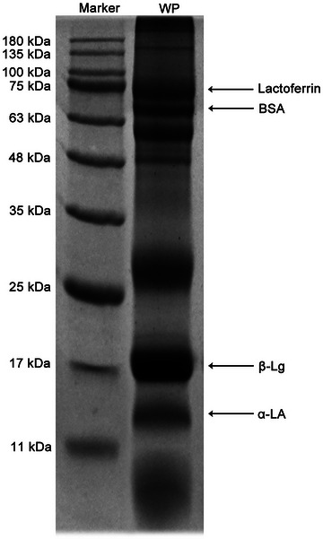
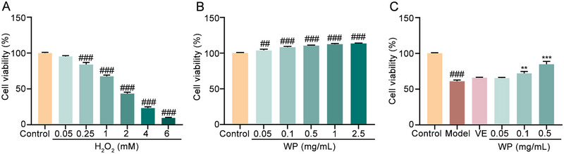
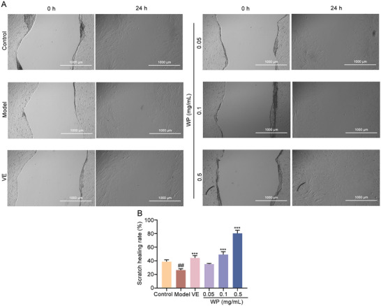
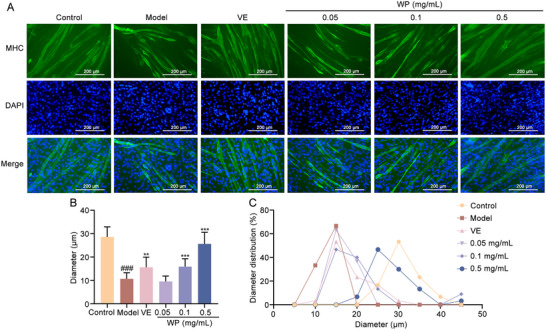
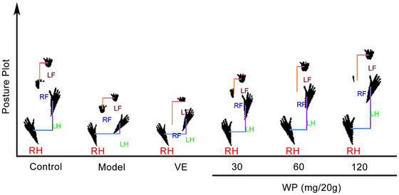
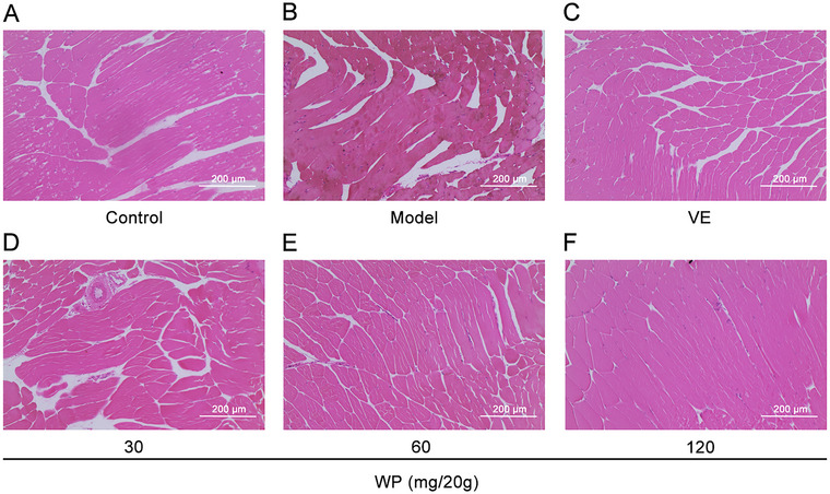
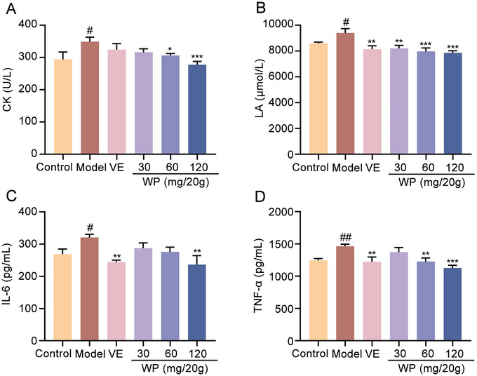
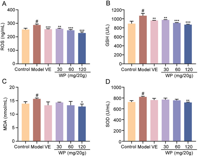
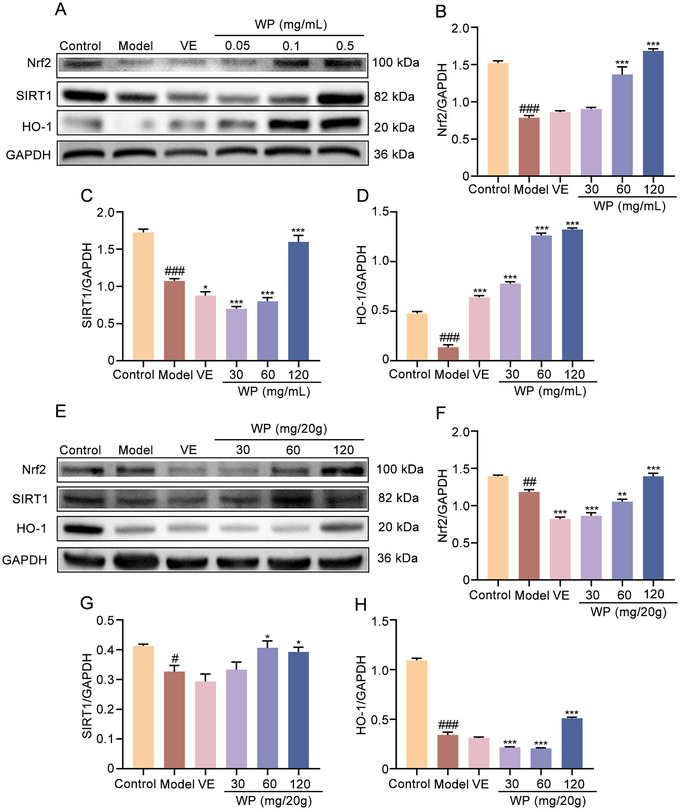
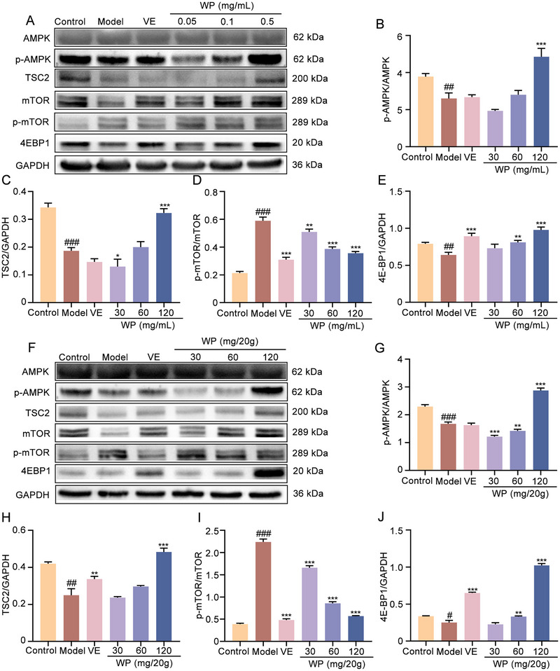
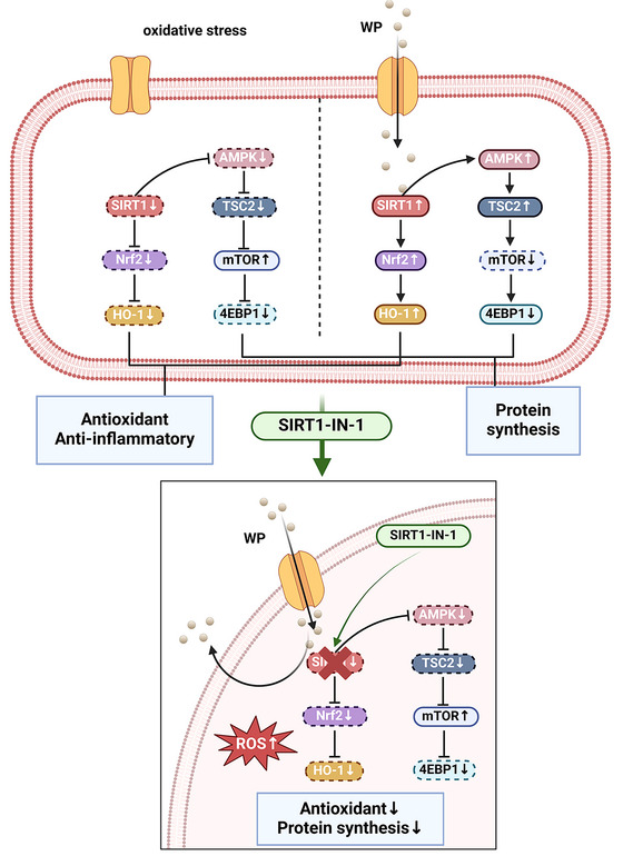
Whey protein (WP) can improve muscle mass and strength. However, its effects and underlying molecular mechanism in promoting recovery from muscle damage caused by excessive physical exercise remains unknown. Therefore, the present study aimed to investigate the therapeutic effect of WP on skeletal muscle injury caused by exogenous oxidants and excessive physical exercise and the potential underlying mechanism. An oxidative stress injury model of mouse skeletal muscle cells was established using hydrogen peroxide (H2O2) and excessive physical exercise. The results revealed that WP effectively improved the migration and differentiation of C2C12 cells exposed to H2O2. Moreover, WP significantly increased the body weight of mice following excessive physical exercise. It also reduced food intake, improved behavioral parameters, enhanced skeletal muscle morphology and function, and promoted protein synthesis, thereby alleviating oxidative stress injury in skeletal muscles. The results further indicated that the mechanism underlying the mitigation of oxidative stress injury in skeletal muscles may involve the silent information regulator sirtuin 1 (SIRT1)/ NF-E2-related factor-2 (Nrf2)/ hemeoxygenase-1 (HO-1) axis. This axis could, in turn, activates the AMP-activated protein kinase (AMPK)/tuberous sclerosis complex subunit 2 (TSC2)/mammalian target of rapamycin (mTOR)/4E-binding protein 1 (4EBP1) pathway, thereby promoting protein synthesis and improving the physiological function of skeletal muscles. This study provides important insights into the role of WP in promoting recovery from muscle damage, offering a basis for future research on WP-based nutritional intervention strategies.

Keywords: AMPK/TSC2/mTOR/4EBP1 pathway; C2C12 cell; Oxidative stress; SIRT1/Nrf2/HO‐1 axis; Skeletal muscle; Whey protein.


Conflict of interest statement

The authors declare no conflicts of interest.

Figures

FIGURE 1
FIGURE 1
SDS‐PAGE pattern of WP. α‐La, α‐lactalbumin; β‐Lg, β‐lactoglobulin; WP, whey protein; SDS‐PAGE, sodium dodecyl sulfate polyacrylamide gel electrophoresis.
FIGURE 2
FIGURE 2
WP antagonized H2O2‐induced oxidative damage in C2C12 cells. (A) Effect of H2O2 on the C2C12 cells viability (one‐way ANOVA). (B) Effect of different WP concentrations on the viability of C2C12 cells (one‐way ANOVA). (C) Effects of WP on C2C12 cells viability after being injured by H2O2 (one‐way ANOVA and t test). Data are expressed as the mean ± SD. ## p < 0.01 and ### p < 0.001 versus control; ## p < 0.01 and *** p < 0.001 versus model. H2O2, Hydrogen peroxide; VE, Vitamin E; WP, whey protein.
FIGURE 3
FIGURE 3
The effect of WP on the migration of C2C12 cells. (A) Effect of WP on the migration of C2C12 cells after 24 h of cell scratching. (B) Effect of WP on scratch healing rate (one‐way ANOVA and t test). Data are expressed as the mean ± SD. ## p < 0.01 versus control; *** p < 0.001 versus model. H2O2, Hydrogen peroxide; VE, Vitamin E; WP, Whey protein.
FIGURE 4
FIGURE 4
The effect of WP on the differentiation of C2C12 cells. (A) MHC immunofluorescence staining of C2C12 cells. (B) The myotube diameter of C2C12 cells (one‐way ANOVA and t test). Data are expressed as the mean ± SD. ### p < 0.001 versus control; ** p < 0.01 and *** p < 0.001 versus model. (C) The diameter distribution of myotubes in C2C12 cells. H2O2, hydrogen peroxide; MHC, myosin heavy chains; VE, Vitamin E; WP, whey protein.
FIGURE 5
FIGURE 5
The effect of WP on gait of mice with exercise injury. LF, left forelimb; LH, left hindlimb; RF, right forelimb; RH, right hindlimb; VE, Vitamin E; WP, whey protein.
FIGURE 6
FIGURE 6
The effect of WP on the morphology and structure of skeletal muscles in exercise injured mice. (A) HE staining of skeletal muscle in normal mice. (B) HE staining of skeletal muscle in mice with exercise injury. (C) HE staining of skeletal muscle in mice with exercise injury under VE intervention. (D–F) HE staining of skeletal muscle in mice with exercise injury under 30 mg/20 g, 60 mg/20 g, 120 mg/20 g WP intervention. VE, Vitamin E; WP, whey protein.
FIGURE 7
FIGURE 7
The effect of WP on the activity of CK, the content of LA and the level of cytokines in the serum of mice with exercise injury. Effects of WP (30, 60, 120 mg/20 g) on serum (A) CK activity, (B) LA content and (C) IL‐6 and (D) TNF‐α levels in mice with exercise injury (one‐way ANOVA and t test). Data are expressed as the mean ± SD. # p < 0.05 and ## p < 0.01 versus control; * p < 0.05, ** p < 0.01 and *** p < 0.001 versus model. CK, creatine kinase; IL‐6, interleukin 6; LA, lactic acid; TNF‐α, tumor necrosis factor alpha; VE, Vitamin E; WP, whey protein.
FIGURE 8
FIGURE 8
The effect of WP on serum antioxidant capacity in mice with exercise injury. The effects of WP (30, 60, 120 mg/20 g) on serum (A) ROS level, (B) GSH and (C) MDA contents and (D) SOD activity in mice with exercise injury (one‐way ANOVA and t test). Data are expressed as the mean ± SD. # p < 0.05 versus control; * p < 0.05, ** p < 0.01 and *** p < 0.001 versus model. GSH, Glutathione; MDA, malondialdehyde; ROS, reactive oxygen species; SOD, superoxide dismutase; VE, Vitamin E; WP, whey protein.
FIGURE 9
FIGURE 9
The effect of WP on SIRT1/Nrf2/HO‐1 axis. (A) WB map of SIRT1/Nrf2/HO‐1 pathway proteins in C2C12 cells induced by H2O2. The effect of WP on the expression of (B) Nrf2, (C) SIRT1 and (D) HO‐1 in C2C12 cells induced by H2O2 (one‐way ANOVA and t test). (E) WB map of SIRT1/Nrf2/HO‐1 pathway proteins in mouse skeletal muscle induced by exercise injury. The effect of WP on the expression of (F) Nrf2, (G) SIRT1 and (H) HO‐1 in mouse skeletal muscle induced by exercise injury (one‐way ANOVA and t test). The samples derive from the same experiment and that gels/blots were processed in parallel. Data are expressed as the mean ± SD. # p < 0.05, ## p < 0.01 and ### p < 0.001 versus control; * p < 0.05, ** p < 0.01 and *** p < 0.001 versus Model. HO‐1, heme oxygenase‐1; Nrf2, NF‐E2‐related factor‐2; SIRT1, silent information regulator sirtuin 1; VE, Vitamin E; WP, whey protein.
FIGURE 10
FIGURE 10
The effect of WP on AMPK/TSC2/mTOR/4EBP1 pathway. (A) WB map of AMPK/TSC2/mTOR/S6K/4EBP1 pathway proteins in C2C12 cells induced by H2O2. Effects of WP on the expression of (B) AMPK, (C) TSC2, (D) mTOR and (E) 4EBP1 in C2C12 cells induced by H2O2 (one‐way ANOVA and t test). (F) WB map of AMPK/TSC2/mTOR/S6K/4EBP1 pathway proteins in mouse skeletal muscle induced by exercise injury. Effects of WP on the expression of (G) AMPK, (H) TSC2, (I) mTOR and (J) 4EBP1 in mouse skeletal muscle induced by exercise injury (one‐way ANOVA and t test). The samples derive from the same experiment and the gels/blots were processed in parallel. Data are expressed as the mean ± SD. # p < 0.05, ## p < 0.01 and ### p < 0.001 versus control; * p < 0.05, ** p < 0.01 and *** p < 0.001 versus model. 4EBP1, 4E‐binding protein 1; AMPK, AMP‐activated protein kinase; mTOR, mechanistic target of rapamycin; TSC2, tuberous sclerosis complex subunit 2; VE, Vitamin E; WP, whey protein.
FIGURE 11
FIGURE 11
Mechanism diagram. SIRT1 may be the center for WP to regulate the antioxidant capacity and protein synthesis of skeletal muscles. (The figure was created using BioRender).

Similar articles

References

    1. Ament, W. , and Verkerke G. J.. 2009. “Exercise and Fatigue.” Sports Medicine (Auckland, N.Z.) 39, no. 5: 389–422. 10.2165/00007256-200939050-00005. - DOI - PubMed
    1. Ataei, L. , Giannaki C. D., Petrou C., and Aphamis G.. 2023. “Effect of Tribulus Terrestris L. Supplementation on Exercise‐Induced Oxidative Stress and Delayed Onset Muscle Soreness Markers: A Pilot Study.” Journal of Dietary Supplements 20, no. 6: 811–831. 10.1080/19390211.2022.2120147. - DOI - PubMed
    1. Bekiroglu, H. , Karaman S., Bozkurt F., and Sagdic O.. 2024. “Characterization of some Physicochemical, Textural, and Antioxidant Properties of Muffins Fortified With Hydrolyzed Whey Protein.” Food Science & Nutrition 12, no. 10: 8105–8117. 10.1002/fsn3.4422. - DOI - PMC - PubMed
    1. Boulier, A. , Denis S., Henry G., et al. 2023. “Casein Structures Differently Affect Postprandial Amino Acid Delivery Through Their Intra‐Gastric Clotting Properties.” Food Chemistry 415: 135779. 10.1016/j.foodchem.2023.135779. - DOI - PubMed
    1. Chang, S. , Wang Z., and An T.. 2024. “T‐Cell Metabolic Reprogramming in Atherosclerosis.” Biomedicines 12, no. 8: 1844. 10.3390/biomedicines12081844. - DOI - PMC - PubMed

MeSH terms

LinkOut - more resources