Case Reports
. 2025 Apr 29:25:e15.
eCollection 2025.

Outcomes of Complex Wound Reconstruction in High-Risk Patients Using Decellularized Extracellular Matrix from Porcine Urinary Bladder

Affiliations
Case Reports

Outcomes of Complex Wound Reconstruction in High-Risk Patients Using Decellularized Extracellular Matrix from Porcine Urinary Bladder

Gracie R Baum et al. Eplasty. .

Abstract

Background: The treatment of complex wounds remains a challenging aspect of reconstructive surgery, given their diverse nature and the frequent need for high-level surgical procedures. Standard treatment with flap coverage can achieve many goals; however, it is not without difficulties, including technical complexity, extended recovery times, donor site morbidity, and vascular complications, particularly in non-optimized patients. Acellular extracellular matrices, such as porcine urinary bladder matrices, have emerged as an alternative approach to support wound healing without the risks of high-level reconstruction. Urinary bladder matrix provides an extracellular matrix scaffold that supports intrinsic tissue regeneration mechanisms, allowing for stable, well-vascularized wound bed formation.

Methods: This retrospective case series examines the outcomes of urinary bladder matrix for the treatment of complex wounds in 21 patients deemed high-risk or unfavorable candidates for surgical management with local or free flap techniques. The patients were treated by 2 surgeons at 2 separate level 1 trauma centers from October 2019 through June 2022. Urinary bladder matrix was the primary wound management modality with serial wound debridement, matrix reapplication, and subsequent wound care tailored to each patient until definitive, stable coverage.

Results: In all cases, urinary bladder matrix facilitated soft tissue remodeling, permitting complete wound re-epithelization or preparation for skin grafting and/or flap coverage. Four patients' wounds re-epithelized on their own, while 17 patients received subsequent skin graft. In 2 of these cases, the initial split-thickness skin graft failed, requiring a second skin graft and or/flap coverage.

Conclusions: Our results demonstrate that urinary bladder matrix facilitates definitive soft tissue reconstruction and can be a valuable adjunct to wound repair, providing a simpler, less morbid treatment option for patients with various comorbidities and injury mechanisms.

Keywords: Acellular Dermis; Extracellular Matrix; Urinary Bladder; Wound Healing; Wounds And Injuries/Therapy.


Conflict of interest statement

Disclosures: Ian L. Valerio is a paid consultant for Integra LifeSciences. All other authors disclose no financial or other conflicts of interest.

Figures
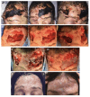
Figure 1
Figure 1
A 38-year-old healthy female involved in a motor vehicle collision sustained a traumatic brain injury and a full-thickness soft tissue injury to the forehead. (A) Initial wound with eschar formation. (B) Following I&D and initial micronized UBM powder application, revealing exposed calvarium with no periosteum and loss of frontalis muscle. (C) Second UBM powder application. (D) Third UBM powder application, covered with a UBM wound matrix sheet. (E) Six-month follow-up showing full re-epithelialization without secondary grafting.
Figure 2
Figure 2
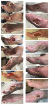
A 64-year-old male with a chronic diabetic foot ulcer over the plantar right foot at the first metatarsal head and plantar great toe with osteomyelitis. The patient underwent amputation of the great toe at the metatarsophalangeal joint, followed by copious I&D. (A) Four days later, a transmetatarsal amputation of the first and second ray was performed. (B, C) Images at 1 and 2 weeks postoperatively. (D) One month later, repeat I&D was performed, closing 14 cm of the wound, leaving a 7 × 3 × 2 cm wound open in the middle of the incision, packed with 500 mg of UBM powder and covered with a 5 × 5 cm UBM sheet matrix bilayer. (E-G) Incremental healing at 10 days, 24 days, and 5 weeks post-UBM application. (H) At 6 weeks post-UBM, a STSG from the proximal thigh was applied. (I, J) Images at 4 days and 7 weeks post-STSG. (K) At 9 weeks post-STSG, the wound size and drainage decreased. (L) At 38 weeks post-STSG, the wound was healed with a central crusted lesion remaining.
Figure 3
Figure 3
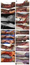
A 40-year-old male sustained a firework injury to the right hand, resulting in a comminuted, open distal radius fracture and carpal bone fractures. (A) Initial I&D with wound VAC placement. (B) One week later, a second I&D and radiocarpal fusion with bone grafting; 500 mg of UBM powder, a 10 × 15-cm UBM 3-layer sheet matrix, and a wound VAC were applied. (C, D) Images at 2 weeks and after a second I&D and UBM application. (E) One week later. (F) Two weeks later, another I&D (11 × 10 × 1 cm), UBM powder, and a 10 × 12-cm sheet of Integra and wound VAC application. (G) Four weeks later, I&D with UBM powder placement over the exposed tendon, followed by STSG and wound VAC. (H) Four weeks post-final UBM placement and STSG. (I) Eight weeks post-final UBM placement and STSG. (J) Wound fully healed at 16 weeks.
Figure 4
Figure 4
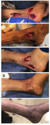
A 56-year-old male involved in an all-terrain vehicle accident sustained a closed left distal tibial shaft fracture. (A) Four weeks after ORIF, the patient presented with a necrotic wound over the lateral ankle fixation site requiring I&D (3 × 10 cm) and wound VAC application. (B) Three weeks later, 500 mg of UBM powder and a 10 × 15-cm UBM 3-layer sheet matrix were applied with wound VAC. (C-E) Images at 4, 6, and 8 weeks post-UBM. (F) At 11 weeks post-UBM, the patient underwent I&D and STSG. (G) One week post-STSG. (H) Five weeks post-STSG (17 weeks post-UBM), the wound was healed with no drainage and a healthy skin graft.
Figure 5
Figure 5
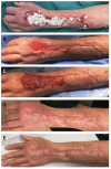
A 43-year-old right-handed male sustained a table saw injury to the right volar palm at the distal crease. (A-C) Initial procedures included 2 revascularization procedures, ORIF of the second metacarpal fracture, digital nerve repair, and tendon repairs. (D, E) Two weeks postoperatively, the wound had central necrosis requiring I&D. The proximal wound was closed with interrupted prolene suture, while the remainder was treated with UBM powder followed by a 5 × 7-cm UBM sheet matrix. (F-I) Weekly UBM powder and sheet applications. (J) Four weeks post-UBM. (K) Six weeks post-UBM, showing near-complete closure without infection or necrosis. (L-O) Healing progression at 9 and 13 weeks. (P) At 5 months, all wounds were healed with restored sensation in the fourth and fifth digits. (Q, R) At 6 months, the patient returned to full manual labor. (S) One-year follow-up showed complete healing, limited scarring, and vascular integrity.
Figure 6
Figure 6
A 54-year-old female with severe combined immunodeficiency and an autoimmune disease on methotrexate presented with a nonhealing surgical wound. (A) After first I&D with UBM powder application. (B) Two weeks later. (C) UBM powder and 3-layer UBM sheet matrix application intraoperatively. (D) Three months later, showing a healthy wound bed. (E) Three years post-treatment.
Figure 7
Figure 7
A 40-year-old male with Crohn disease developed a fistula with necrotizing infection involving the abdomen and right leg. After multiple surgical debridements, the wound was treated in a single stage with UBM powder over the knee and posterior exposed tendon, followed by a single-layer Integra matrix and STSG. One-year follow-up showed wounds healed within 6 to 8 weeks.
Figure 8
Figure 8
A 27-year-old male sustained a left lower extremity blast-related trauma with a Gustilo-Anderson type 3B tibial fracture and extensive periosteal loss. (A) Fully debrided wound exposing the tibia. (B) Exposed tibia with absent periosteum. (C) Identification of vessel loops before reconstruction. (D) Hybrid reconstruction using latissimus dorsi free flap for superior coverage, serial UBM application, and Integra bilayer for the distal third of the tibia. (E) Initial post-reconstruction. (F) Four weeks post-reconstruction with well-vascularized tissue. (G) Four weeks post-STSG application, demonstrating coverage over hybrid reconstruction with serial UBM and Integra placement.
Figure 9
Figure 9
A 68-year-old male with a left lateral ankle wound due to a diabetic ulcer. (A, B) Initial wound with I&D and application of UBM powder and a 3-layer UBM sheet matrix. (C) Following the first application, granulation tissue formation covered exposed bone. A second UBM application was performed. (D) Repeat I&D and UBM placement with subsequent skin grafting. (E) The skin graft integrated around the wound margin but failed centrally. (F) The central wound was re-grafted 2 weeks later. (G) UBM powder was applied in the clinic 2 to 4 weeks later. (H) One month post-UBM powder application. (I) Two-year follow-up demonstrating complete epithelialization with no exposed bone.
Figure 10
Figure 10
A 67-year-old male with a venous stasis ulcer leading to an open left ankle wound with exposed medial malleolus. (A) Serial debridement to prepare the wound. (B) The proximal wound was closed, and UBM powder and a UBM sheet matrix were applied to the remaining wounds. (C) Two weeks post-UBM, additional UBM powder was applied. (D) One month post-UBM. (E) Two-month follow-up demonstrating full epithelialization.
Figure 11
Figure 11
A 72-year-old male with a superficial sternal wound infection 3.5 months after coronary artery bypass grafting (CABG), complicated by postoperative Clostridioides difficile infection. Despite a 2-week antibiotic course, the infection progressed, requiring 3 I&Ds before applying 1500 mg UBM powder and a 10 × 15-cm UBM burn matrix, followed by a wound vac. (A) One-week post-UBM. (B) Two weeks post-UBM. (C) Three weeks post-UBM with I&D. (D) Five weeks post-UBM. (E) Six weeks post-UBM. (F) Four weeks later, superficial debridement was performed, and a 3 × 5-cm STSG was placed over the sternal wound. (G) Two weeks post-STSG. (H) Five months post-UBM with diffuse scabbing. (I-K) Fourteen weeks later, bilateral pectoralis major advancement with anterior chest wall skin flap mobilization was performed. (L) At 11 months post-UBM, the incision was well healed, and the patient resumed full activity, including pushups, without pain.
Figure 12
Figure 12
A 33-year-old male with uncontrolled diabetes, polyneuropathy, and chronic multifocal osteomyelitis developed a chronic left wrist and palm infection with multiple draining abscesses. (A) Serial I&D procedures followed by 500 mg UBM powder and bilayer UBM sheet matrix placement. (B) Eighteen days, (C) 3 weeks, (D) 4 weeks, and (E) 6 weeks post-UBM application showing progressive wound healing. (F) Nine weeks post-UBM with granulation tissue formation but persistent areas of exposed tendon. Additional UBM powder was applied, followed by an STSG and wound vac. (G) One week, (H) 5 weeks, and (I) 12 weeks post-STSG demonstrating progressive wound closure.
Figure 13
Figure 13
An 88-year-old male with multiple comorbidities sustained a gunshot wound to the left hand, resulting in a large dorsal soft tissue defect and an open comminuted second metacarpal fracture with segmental bone loss. (A) One week post-ORIF and I&D with Integra placement and a wound vac. (B) One month, (C) 2 months post-ORIF. (D) Repeat I&D with 200 mg UBM powder and a 5 × 5-cm UBM sheet matrix application. (E) One week and (F) 1 month post-UBM. (G) Three weeks post-I&D with STSG and subcutaneous fat grafting. (H) Three-month post-STSG. (I) Seven to 8 months post-STSG, with full healing. (J) One-year and (K) 13-month follow-up demonstrating durable wound closure.
Figure 14
Figure 14
A 52-year-old male sustained a crush injury to the right leg from a 1000-pound air conditioning unit. (A) Initial I&D and external fixation followed by ORIF of the tibia with an intramedullary nail. (B) A bony defect was filled with an orthobiologic graft, and gastrocnemius and soleus rotational flaps were performed. (C) Two and a half weeks later, necrosis of the soleus flap was noted; UBM powder was applied with a wound vac. (D) Two weeks post-UBM, an STSG was placed. (E) Four weeks post-UBM, additional debridement was required. UBM powder and a 10 × 15-cm UBM sheet were placed over the wound. (F) One week post-UBM. (G) Three weeks post-UBM with additional STSG. (H) Two weeks, (I) 4 weeks, and (J) 5 weeks post-STSG. (K) A small area of exposed bone was debrided, and UBM powder was applied. (L) Six weeks and (M) 11 weeks post-final UBM application, demonstrating wound closure.
Figure 15
Figure 15
A 33-year-old male sustained a gunshot wound to the right mid-tibia, resulting in a mangling limb injury with a type 3B open tibial fracture and a 120-mm tibial nerve gap. (A) Initial I&D with external fixation, followed by ORIF with 1 g UBM powder and a 10 × 15-cm UBM sheet matrix application. (B) Eight days post-injury, the wound showed granulation tissue formation. An STSG and tibial nerve allograft repair were performed. (C) Three, (D) 8, and (E) 12 days post-STSG. (F) Six weeks post-STSG, with additional UBM powder and wound vac placement. (G) Five days post-UBM, additional STSG was performed. (H) Ten days post-STSG. (I) Three weeks later, I&D and another UBM application were performed. (J) Six weeks post-STSG, with partial flap loss. (K) A gracilis free flap was used for soft tissue coverage. (L) Four days post-flap. Also see Video 2.
Figure 16
Figure 16
A 57-year-old male with poorly controlled diabetes sustained a closed left trimalleolar ankle fracture after a fall. (A) Seven weeks post-injury, a 10 × 3.5 cm open wound with exposed hardware was debrided and treated with a wound vac. Three days later, a 10 × 4 cm full-thickness wound was treated with 20 mg UBM powder and a 10 × 7 cm UBM sheet matrix. (B) Twelve weeks post-UBM, repeat I&D and STSG were performed. (C) Four months later, the wound showed re-epithelialization with no signs of infection. (D) Eleven-week follow-up demonstrating continued wound improvement with only minor central scabs.

References

    1. Park H, Copeland C, Henry S, Barbul A. Complex wounds and their management. Surg Clin North Am. Dec 2010;90(6):1181-1194. doi:10.1016/j.suc.2010.08.001 10.1016/j.suc.2010.08.001 - DOI - DOI - PubMed
    1. Lee CK, Hansen SL. Management of acute wounds. Surg Clin North Am. Jun 2009;89(3):659-676. doi:10.1016/j.suc.2009.03.005 10.1016/j.suc.2009.03.005 - DOI - DOI - PubMed
    1. Beyene RT, Derryberry SL, Barbul A. The effect of comorbidities on wound healing. Surg Clin North Am. 2020. Aug;100(4):695-705. doi:https://doi.org/10.1016/j.suc.2020.05.002 10.1016/j.suc.2020.05.002 - DOI - DOI - PubMed
    1. Janis JE, Kwon RK, Attinger CE. The new reconstructive ladder: modifications to the traditional model. Plast Reconstr Surg. Jan 2011;127 Suppl 1:205s-212s. doi:10.1097/PRS.0b013e318201271c 10.1097/PRS.0b013e318201271c - DOI - DOI - PubMed
    1. Boyce DE, Shokrollahi K. Reconstructive surgery. BMJ. 2006;332(7543):710-712. doi:10.1136/bmj.332.7543.710 10.1136/bmj.332.7543.710 - DOI - DOI - PMC - PubMed

Publication types

LinkOut - more resources